History and exam
Key diagnostic factors
common
exposure to an orthoebolavirus in previous 21 days
Human-to-human transmission occurs via contact with body fluids (e.g., sweat, blood, feces, vomit, saliva, genital secretions [including semen], amniotic fluid, and breast milk) from infected patients.[43][44] Virus levels in these fluids are particularly high in more severe or advanced infection. The incubation period after infection is 2-21 days.[3] Incubation periods may be shorter in children.[76]
Contacts of infected patients (including healthcare workers and household contacts) are at risk of infection if the person was exposed to body fluids of the infected patient without appropriate protective equipment. Household contacts of infected patients are at a higher risk of infection if there is active diarrhea, vomiting, or bleeding.[43]
Body fluids remain infectious even after death. As a consequence, many infections have occurred at traditional funeral services in Africa where mourners touch the bodies of the deceased. Super-spreading events in the community are also increasingly recognized as a contributing factor: a funeral of a traditional healer in Sierra Leone in 2015 was linked to 300 cases.[47] In one study, it was found that super-spreaders were responsible for approximately 61% of infections in the 2014 outbreak.[48]
People who have traveled to endemic areas are considered to be at high risk of infection. Up-to-date knowledge of the geographical locations of active epidemics helps to clarify the patient’s epidemiologic risk.
fever
Presenting symptom in up to 90% of patients, and is often >102.2°F (39.0°C) with a remitting pattern.[18][106] Some patients may initially have a low-grade fever with no other symptoms, or alternatively the temperature may be near normal at first evaluation.[108]
The temperature threshold for fever differs among countries and guidelines, and using a lower temperature threshold (e.g., ≥99.5°F [37.5°C]) increases the sensitivity of finding cases.[106][109] The World Health Organization use a threshold of >100.4°F (38°C).[110] However, in a large cohort in Sierra Leone, <30% had a fever of ≥100.4°F (38°C) at presentation, although a history of fever was reported by 89% of patients.[22] Another study in Sierra Leone found that 25% of children did not have a history of fever, or a temperature ≥100.4°F (38°C) at the time of admission.[105]
Reported in 76% of patients in the 2014 outbreak.[100]
Presence is enough to raise concern for infection in the appropriate epidemiologic context.
Wide variations in body temperature can be observed, especially in children.[105][107] Patients are often normothermic or hypothermic in the later stages of fatal infection.[16][17][102]
myalgia
Other diagnostic factors
common
fatigue
anorexia
Reported in 64% of patients in the 2014 outbreak.[100]
diarrhea
Common feature of infection, present in 88% of patients in a previous outbreak.[17]
Reported in 51% of patients in the 2014 outbreak.[18][100]
May be bloody.
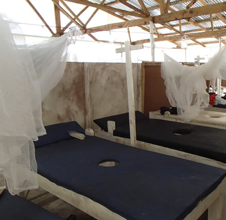
Cholera beds may be used for cases of profuse diarrhea in undeveloped countries.[Figure caption and citation for the preceding image starts]: Cholera beds with central hole in mattress to manage patients with profuse diarrhea at an Ebola treatment center in West Africa, 2014From the personal collection of Catherine F. Houlihan, MSc, MB ChB, MRCP, DTMH; used with permission [Citation ends].
vomiting
severe headache
abdominal pain or heartburn
Reported in 40% of patients in the 2014 outbreak.[100]
It may be difficult to distinguish heartburn from lower anterior chest pain or dysphagia. Dysphagia and heartburn are likely due to esophagitis.
cough, dyspnea, chest pain
Chest pain and cough reported in 10% and 7% of patients respectively in the 2014 outbreak; however, direct involvement of the lungs has only rarely been reported.[19][111]
Difficulty breathing reported in 18% of patients in the 2014 outbreak.[100]
Respiratory symptoms tend to be more common in children compared with adults; however, data are limited.[102][103] Difficulty breathing was reported in 14% of children in the 2014 outbreak.[105]
sore throat
prostration
Profound prostration is a typical finding reported in 73% of patients in the 2014 outbreak.[130]
uncommon
maculopapular rash
Developed early in the course of illness in approximately 25% to 52% of patients in previous outbreaks.[16]
Reported in 3% of patients in the 2014 outbreak.[100]
Frequently described as nonpruritic, erythematous, and maculopapular. It may begin focally, then become diffuse, generalized, and confluent. Some have described it as morbilliform. May become purpuric or petechial later on in the infection in patients with coagulopathy.[23]
May be difficult to discern in dark-skinned patients.
bleeding
Presence suggests advanced infection and presence of disseminated intravascular coagulation.
Bleeding manifestations (e.g., epistaxis, bleeding gums, hemoptysis, easy bruising, conjunctival bleeding, hematuria, oozing from injection or venipuncture sites) were present in 30% to 36% of infected patients in previous outbreaks; however, they were reported in only 11% of patients in the 2014 outbreak.[8][16][17][100]
Massive bleeding is usually only observed in fatal cases, and typically occurs in the gastrointestinal tract (e.g., melena, bloody diarrhea).[16] In a previous outbreak, melena was present in 8% of fatal infections and 16% of survivors.[17]
Internal bleeding may be missed if there are no external signs.
Bleeding manifestations are less common in children.[104]
hepatomegaly
In a previous outbreak, tender hepatomegaly with the edge of the liver palpable below the ribcage was present in 2% of fatal infections and 8% of survivors.[17]
lymphadenopathy
Enlarged lymph nodes have been reported.[16]
hiccups
Sign of advanced infection and poor prognosis, typically seen in the last 2-3 days of fatal infections.[16]
May be due to uremia, hypokalemia, hyponatremia, hypocalcemia, or hypocarbia due to respiratory compensation of metabolic acidosis.
In a previous outbreak, hiccups were present in 17% of fatal infections and 5% of survivors.[17]
Reported in 13% of patients in the 2014 outbreak.[100]
tachycardia
May be seen in the later stages of fatal infections.[16]
hypotension
Feature of preterminal disease and shock. It is under-documented in field studies owing to a lack of measuring equipment in endemic areas.[16]
However, septic shock with vascular leakage and microcirculatory failure does not appear to be a dominant feature.
neurologic signs
Confusion was reported in 9% of patients in the 2014 outbreak.[100] It appeared to be more common compared with previous outbreaks, and is a predictor of death.[105][107][130] Confusion may be multifactorial in children and is associated with a poor prognosis.[105][107]
Often coexist with bleeding and hypotension making fluid resuscitation hazardous.
Encephalopathy is possibly related to electrolyte disturbances, uremia, and cerebral hypoperfuson in terminal infection.
Seizures occurred in 2% of fatal infections in a previous outbreak.[17]
Risk factors
strong
living or working in, or arrival from, endemic area in previous 21 days
People living or working in endemic areas (e.g., West Africa, Democratic Republic of the Congo) are at high risk of infection. However, recent arrival from endemic areas is also a significant risk factor. Most patients with suspected infection in developed countries will be returning travelers and healthcare workers who have cared for patients during outbreaks.
Up-to-date knowledge of the geographic locations of active epidemics helps to clarify the patient’s epidemiologic risk.
contact with infected body fluids
Human-to-human transmission occurs via contact with body fluids (e.g., sweat, blood, feces, vomit, saliva, genital secretions [including semen], amniotic fluid, and breast milk) from infected patients, or objects contaminated with infected body fluids.[43][44] Virus levels in these fluids are particularly high in more severe or advanced infection. The incubation period after infection is 2-21 days.[3] Incubation periods may be shorter in children.[76]
Contacts of infected patients (including healthcare workers and household contacts) are at risk of infection if the person was exposed to body fluids of the infected patient without appropriate protective equipment. Household contacts of infected patients are at a higher risk of infection if there is active diarrhea, vomiting, or bleeding.[43]
Body fluids remain infectious even after death. As a consequence, many infections have occurred at traditional funeral services in Africa where mourners touch the bodies of the deceased.[77] Super-spreading events in the community are also increasingly recognized as a contributing factor: a funeral of a traditional healer in Sierra Leone in 2015 was linked to 300 cases.[47] In one study, it was found that super-spreaders were responsible for approximately 61% of infections in the 2014 outbreak.[48]
Sexual transmission has been documented during active infection. The virus can still be detected in semen more than 12 months after recovery from infection, possibly due to testicular tissue being an immunologically-protected site.[51] This means that sexual transmission may be possible long after the infection has resolved, and such cases were confirmed during the 2014 outbreak.[44][49][50][52][53][54][55][56]
occupational exposure
Healthcare workers in contact with infected patients without proper protective equipment are at high risk, and most epidemics have resulted in numerous infections in healthcare professionals.
Needlestick injuries from an infected donor are a very high-risk exposure depending on the inoculum and nature of the injury. Use of nonsterile needles was responsible for the nosocomial spread of the first epidemic in 1976.[24] Accidental needle exposure has occurred in research laboratories in the UK, Russia, and Germany. The incubation periods in such cases may be considerably shorter compared with human-to-human transmission.[7][17][49]
Other high-risk occupations include those where people work with primates or bats from endemic areas, or high-risk clinical samples.
butchering or consumption of meat from infected (or potentially infected) animals
This route of transmission is likely to be a cause of animal-to-human transmission in sporadic epidemics.[78]
Use of this content is subject to our disclaimer
