
Etiology
Hantaviruses are a group of negative-stranded RNA viruses in the family Hantaviridae.[3][7]
Hantavirus cardiopulmonary syndrome (HCPS) usually results from exposure to rodent excreta containing one of the identified pathogenic hantaviruses. In the US, Sin Nombre virus (SNV), Bayou virus (BAYV), Black Creek Canal virus (BCCV), New York virus (NYV), and Monongahela virus (MONV) are the most common causes. Other hantaviruses are the cause of HCPS in Central and South America (e.g., most commonly Andes virus [ANDV] in South America).[3]
Early in the recognition of HCPS, identification of the causative virus was determined from patient isolates and isolates from deer mice captured in and around the patients' dwellings.[22] Hantaviruses have coevolved with their specific rodent hosts, are asymptomatic in the rodent host, and are transmitted intraspecies through biting and salivary exposure. Hantavirus infections are predominantly transmitted to humans through incidental contact with rodent urine or saliva.[23] Aerosol, mucous membrane, and nonintact skin exposure to rodent excreta, rodent bite, and laboratory accidents are all possible routes of exposure. Viability of the hantaviruses is 2 to 3 days at normal room temperature. Exposure to sunlight will decrease the time of viability.[Figure caption and citation for the preceding image starts]: Peromyscus maniculatus: the deer mouse, vector for Sin Nombre virus (SNV), which causes most cases of hantavirus cardiopulmonary syndromeCDC Public Health Image Library (PHIL), James Gathany [Citation ends].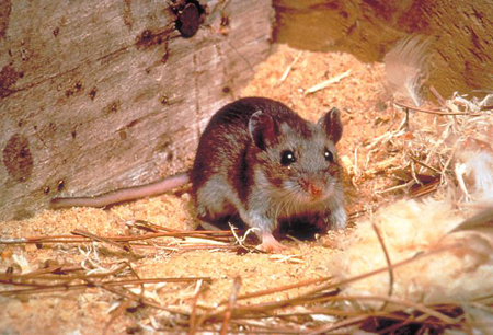
Human-to-human transmission
Human-to-human transmission has only been reported with ANDV.[24]
In North America, no human-to-human transmission of hantaviruses has been demonstrated.
The outcome of 5 cases of SNV infection in pregnancy included 1 maternal death and 2 fetal losses. Exam of 2 fetal autopsies and 3 placentas, and serologic follow-up of the 3 surviving children, did not show evidence of perinatal transmission of SNV.[25]
In South America, ANDV is a major cause of HCPS and is most frequently acquired by contact with or through aerosols of excreta and secretions of infected rodents.[26] However, cases of human-to-human transmission of ANDV have been reported from territories of Argentina and Chile.[27][28]
In Chile, human-to-human transmission was documented to occur mainly in family clusters.[27][28]
In a prospective study, sexual partners of an infected person had a 10-fold increased risk of infection compared with other household contacts.[29]
Close contact with a sick person during the prodromal phase of the disease (12-27 days from initial exposure of the source case) appeared to increase the chances of human-to-human transmission.[4]
Human-to-human transmission of ANDV was reported during the 2026 M/V Hondius cruise ship outbreak.[21]
Transmission was likely amplified by various factors including the older age of people on the ship (mean age 65 years) and a substantial proportion of people having comorbidities, as well as crowded living spaces and exposure to shared indoor spaces.[30]
Several studies of patients infected with ANDV have shown the presence of the virus in different body fluids such as blood, respiratory secretions, gingival crevicular fluid, saliva, and urine. These observations might explain in part why it is possible to contract the virus by close contact with any of these fluids from a patient in the prodromal phase of illness.[31]
Although there are no studies to confirm the route of transmission, possible transmission routes may include sexual transmission or other direct contact, direct contact via breastfeeding, or respiratory. No studies ruled out respiratory or fomite transmission.[24]
A meta analysis of the available evidence does not support routine human-to-human transmission of ANDV.[32]
Nosocomial transmission of ANDV has been reported in healthcare workers in the setting of delayed implementation of transmission-based precautions or breaches in infection prevention and control measures.[28][33]
Prior seroprevalence studies conducted in Chile with healthcare personnel working at hospitals where patients with ANDV infection were treated showed the presence of anti-ANDV immunoglobulin G antibodies in healthcare workers was similar to that of the general population.[34]
There is no evidence of asymptomatic or presymptomatic transmission.[24]
Pathophysiology
Following inhalation exposure to hantavirus in rodent excreta, there is an incubation period of 9 to 33 days (median 14 to 17 days, and up to 3 weeks after rodent bite), during which the virus replicates in pulmonary macrophages and dendritic cells without causing cell death and is distributed to lymphoid organs.[35][36][37]
In human-to-human transmission caused by ANDV, the reported incubation period ranged from 9 to 40 days (mean 21.6 to 27.5 days) across studies. The reported serial interval ranged from 4 to 40 days (mean 19.6 to 25.7 days).[24]
After the incubation period, viremia is produced that infects target endothelial cells and stimulates T cells. Host neutralizing antibody is produced that may mitigate the severity of the infection.[38] Immune-stimulated T cells are distributed to areas of hantavirus concentration, particularly the lung and heart interstitium.[39] A nonspecific viral syndrome with predominantly severe myalgias and gastrointestinal symptoms follows that is often not recognized. Rarely, the illness does not progress; however, usually patients are admitted to the hospital within 5 days having progressive respiratory failure with pulmonary edema, metabolic acidosis, and cardiogenic shock.[40]
Progressive disease results from infected endothelial cell function being disturbed and local T-cell cytokine production, which causes capillary leak in target organs.[41][42] The capillary leak may be the result of pathogenic hantaviruses using an alpha-variable, beta-3 integrin ligand on the endothelial cell and platelet surfaces, resulting in disturbed endothelial cell migration and endothelial cell barrier function.[37][43]
Studies in a Syrian hamster hantavirus pulmonary syndrome (HPS) model have suggested a direct effect of hantavirus on resting platelets through binding to the platelet beta-3 integrin with resultant platelet cross-linking and clumping on endothelial surfaces. There is also binding to the beta-3 integrin on the resting endothelial cells with disruption of vascular endothelial growth factor receptor activity, increased phosphorylation and internalization of vascular endothelial-cadherin, and loss of cellular tight junction competence.[44]
All pathogenic hantaviruses have also been demonstrated to have an immunoreceptor tyrosine-based activation motif on their G1 envelope glycoproteins, which may modulate cellular downstream signaling and endothelial and immune cell function.[45] Sensitized mononuclear cells infiltrate the lung, myocardial interstitium, and spleen to produce cytokines, particularly tumor necrosis factor-alpha and interferon-gamma, resulting in pulmonary edema and myocarditis.[46][47] Hantaviruses may also attach to beta-2 integrin receptors on neutrophils and induce the release of neutrophil extracellular traps.[48]
Clasificación
Virus taxonomy[3]
Hantaviruses belong to the family Hantaviridae, genus Orthohantavirus, and order Bunyavirales. New World hantaviruses are associated with HCPS.
Clinically significant species identified in the US and associated with HCPS include:
Sin Nombre orthohantavirus (also known as Orthohantavirus sinnombreense)
Sin Nombre virus (SNV)
New York virus (NYV)
Monongahela virus (MONV)
Bayou orthohantavirus (also known as Orthohantavirus bayoui)
Bayou virus (BAYV)
Black Creek Canal orthohantavirus (also known as Orthohantavirus nigrorivense)
Black creek canal virus (BCCV)
Clinically significant species identified in Central and South America and associated with HCPS include:
Andes orthohantavirus (also known as Orthohantavirus andesense)
Andes virus (ANDV)
Castelo dos Sonhos (CASV)
Lechiguanas virus (LECV or LECHV)
Oran virus (ORNV)
Laguna Negra orthohantavirus (also known as Orthohantavirus negraense)
Laguna Negra virus (LANV)
Maripa virus (MARV)
Rio Mamore virus (RIOMV)
Choclo orthohantavirus (also known as Orthohantavirus chocloense)
Choclo virus (CHOV)
Unclassified orthohantaviruses associated with HCPS include:
Anajatuba virus (ANJV)
Araraquara virus (ARQV)
Araucaria virus (ARAUV)
Bermejo virus (BMJV)
Juquitiba virus (JUQV)
Maciel virus (MACV)
Paranoá virus (PARV)
Pergamino virus (PERV)
Based on phylogenetic studies, the South American hantavirus strains have been separated into three monophyletic clades: Andes, Laguna Negra, and Rio Mamore. Each of these clades has been classified as a unique species. The Andes clade is found in Argentina, Bolivia, Brazil, Chile, Paraguay, and Uruguay. The Laguna Negra clade is found in Argentina, Bolivia, Brazil, and Paraguay. The Rio Mamore clade is found in Brazil, Bolivia, French Guyana, Paraguay, and Peru.[4]
内容使用需遵循免责声明