Approach
Marburg virus infection is a notifiable disease. The case definition for Marburg virus infection is broad and includes a long list of possible differential diagnoses.
The initial assessment of a patient with suspected Marburg virus infection hinges on two main factors:[19][20]
Epidemiological risk (e.g., living or working in, or travel to, an endemic area in previous 21 days; or laboratory exposure); and
Presence or history of a high fever.
Infection prevention and control
Infection prevention and control (IPC) is of immediate concern and local protocols should be followed. As soon as a clinician decides Marburg virus disease is a possible diagnosis for their patient’s illness, high-level precautionary isolation procedures and personal protective equipment (PPE) should be used until the infection is either confirmed or excluded. It is extremely important to minimise the risk of transmission while working up the patient.[19][21] Although 'wet symptoms' such as vomiting/diarrhoea may increase transmission risk, Marburg virus should be considered highly infectious and high-level precautions should be used regardless of symptoms at presentation.
The World Health Organization (WHO) recommends the following IPC principles in healthcare settings.[22]
An IPC ring approach is recommended in healthcare facilities and communities during the management of cases.
In geographic areas where the virus is circulating, all people should be screened at the first point of contact with a healthcare facility, using a no-touch technique, to enable early recognition of suspected cases.
All suspected cases should be triaged to determine the severity of disease and identify patients in need of immediate care.
Patients with suspected or confirmed infection should be isolated, preferably in a single room.
Interaction with family and visitors should be facilitated to promote wellbeing, while preventing direct contact with others.
Hand hygiene should be performed using an alcohol-based hand rub or soap and running water using the correct technique.
Bleach/chlorine solutions may be used in emergency situations when these methods are not available until they become available.
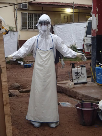
Appropriate PPE should be worn when in contact with a suspected or confirmed case.
Mucous membranes of the eyes, mouth, and nose should be completely covered. A face shield or goggles (under the head-and-neck covering) should be used. Fluid-resistant surgical or medical masks with a structured design (so that they do not collapse against the mouth) are recommended. A fluid-resistant particulate respirator should be used during aerosol-generating procedures.
Gloves and a disposable gown (or coverall) and apron made of fabric that has been tested for resistance to penetration by body fluids or bloodborne pathogens should be worn. Nitrile gloves are preferred over latex gloves.
Specific PPE requirements depend on the level of patient contact (i.e., indirect contact such as screening and triage vs. direct contact of a case). PPE is not required during screening activities where a distance of at least 1 metre can be guaranteed and a no-touch approach is strictly followed.
Surfaces should be disinfected (using the wiping method) in facilities and settings that provide care to patients with suspected or confirmed infection.
All waste generated from the care of a patient with suspected or confirmed infection should be treated as infectious waste.
Heavily-soiled linens from patients should be disposed of safely (e.g., incinerated).
Healthcare workers with occupational exposure should be immediately assessed for exposure risk and managed accordingly.
The WHO suggests exclusion from work for 21 days.
More detailed IPC guidance is available from the WHO:
Guidance is also available from the Centers for Disease Control and Prevention (CDC):
[Figure caption and citation for the preceding image starts]: Healthcare worker in personal protective equipment at an Ebola treatment centre in Sierra Leone, 2014From the personal collection of Chris Lane, MSc (Public Health England/World Health Organization); used with permission [Citation ends].
History
A detailed history can clarify the risk for Marburg virus infection and other acute febrile syndromes.
People living or working in endemic areas (Central and Eastern Africa) or working with Marburg virus or virus-carrying monkeys in a laboratory setting are at the highest risk of infection.[23] Recent arrival (within 21 days) from endemic areas is also an important risk factor. Visiting caves or mines inhabited by African fruit bat (Rousettus aegyptiacus) colonies increases suspicion of Marburg virus infection in symptomatic patients.[13]
In developed countries, most patients with suspected infection will be returning travellers, healthcare workers who have cared for patients during outbreaks, or laboratory workers with occupational exposure. Therefore, a comprehensive history of travel, work, and attendance of burials or funerals is extremely important. Knowledge of the geographical locations of active or prior epidemics helps to clarify the patient's epidemiological risk.
High-risk occupations include those where people work with primates or bats from endemic areas or with high-risk clinical samples, and healthcare or laboratory workers in endemic or outbreak settings.
As malaria is still the most common cause of febrile illness in returning travellers from Africa, the presence of risk factors for acquiring malaria should be assessed (e.g., living/working in, or travelling to, endemic area; inadequate or absent chemoprophylaxis; not using insecticides or bed nets).[24] However, co-infection with malaria and viral haemorrhagic fever does occur, and known or suspected diagnosis of malaria does not exclude a possible diagnosis of Marburg virus infection.[25]
Exposure risk
Contacts of infected patients (including healthcare workers and household contacts) are at risk of infection if the person was exposed to body fluids of the infected patient without appropriate protective equipment. The incubation period after infection is typically 5 to 10 days.[1][13] Brief interactions, such as walking by an infected person or through a hospital, do not constitute close contact.
Contact is defined by the WHO as someone who has:[26]
Slept in the same household as a patient
Had direct physical contact with the patient during the illness or at the funeral
Touched the patient's body fluids or clothes/bed linens during the illness or soon after death, including washing clothes soiled during the illness
Been breast-fed by the patient (babies).
Other infection risks include exposure to dead or sick animals, caves, or mines in endemic areas, and laboratory contacts.[26] In endemic areas, viral transmission may occur from animals to humans even without a notable bite or scratch.[27]
Case definitions for Ebola virus and Marburg virus can be found on the WHO website:
Symptoms
The incubation period after infection is typically 5 to 10 days, although some estimates range from 2 to 26 days.[12][13][14] Patients are not considered infectious until they develop symptoms.
The initial presentation is non-specific, which makes early clinical diagnosis difficult; however, typical symptoms include:[1][13]
High fever
Chills
Headache
Myalgia and malaise
Nausea/vomiting
Diarrhoea
Abdominal pain
Myalgia
Prostration
Sore throat
Hiccups
Difficulty breathing
Unexplained bruising or bleeding.
Three phases of illness are typically recognised, starting with a few days of high fever, headache, myalgia, and malaise, and followed by a gastrointestinal phase where anorexia, diarrhoea, vomiting, abdominal pain, and dehydration are prominent.[13] Other symptoms may include conjunctivitis and rash on the mucous membranes.[13] In the second phase, the patient may either recover, or deteriorate with a third phase of illness which includes collapse, neurological manifestations, and bleeding and generalised rash in some patients. Fatalities often occur during this phase, approximately day 8 to 16 of illness.[14][28]
Physical examination
A full physical examination should be undertaken with the aim of excluding a clear alternative diagnosis while looking for signs of viral haemorrhagic fever (e.g., conjunctival injection, purpuric rash, or other signs of bleeding). Note that Marburg virus disease is a multi-phase illness first presenting with fever and non-specific symptoms, and later progressing to include febrile gastrointestinal symptoms. It is important to note that not all patients will have signs or symptoms of bleeding or coagulopathy.[13][28]
Vital signs should be taken:
Fever: the presenting symptom in most patients, its presence is enough to raise concern for infection in the appropriate epidemiological context. Wide variations in body temperature can be observed during the course of illness with normothermia or hypothermia occurring in the later stages of fatal infection. High fever (>40°C [104°F]) is common.[13][14]
Blood pressure: hypotension is a feature of dehydration and shock and is present in later-stage disease.[13] It is under-documented in field studies owing to a lack of measuring equipment in endemic areas.[14]
Pulse rate: bradycardia may be present in the initial stages of illness; however, tachycardia may be seen in the later stages of infection.[14]
Respiratory rate: tachypnoea may be present in later stages of illness, resulting from metabolic acidosis.[27]
Initial investigations
All specimens should be collected according to the same strict protocols as those for Ebola virus. The CDC and WHO have published guidance on this:
The main confirmatory test for Marburg virus infection is a positive reverse transcriptase polymerase chain reaction (RT-PCR) for Marburg virus from blood or oral (buccal membrane) swab.[13] This test should be ordered in all patients with suspected Marburg infection while the patient is in isolation. It has the advantage of returning a result 4 to 48 hours before ELISA testing.[29] In high-resource settings, the test may only be available in regional or national laboratories which have biosafety level 4 facilities.[30] Viral RNA can be detected in the patient's blood by RT-PCR from as early as day 1 up to days 6 to 17 of symptom onset.[29][31] A positive PCR result implies that the patient is potentially infectious, particularly in the presence of active diarrhoea, vomiting, or bleeding. If negative, the test should be repeated within 48 hours since viral load is low and can be undetectable early in the course of the illness. Negative tests should be repeated at least 72 hours into infection to rule out a diagnosis if it is strongly suspected (or confirm resolution of infection).[29][31] It is likely that higher viral load correlates with adverse outcomes and increased mortality.[32]
Malaria is still the most common cause of fever in people who live, work, or travel to endemic areas.[24] All people visiting a malarial area within the last one year should be tested for malaria and treated empirically for malaria if filovirus disease is suspected.[25] In the case of a positive rapid diagnostic test result for malaria, the infection should be treated while continuing to assess the risk for Ebola or Marburg virus infection and noting the possibility of dual infection.
If Ebola or Marburg virus infection is suspected, it is recommended that appropriate confirmatory tests for Ebola and/or Marburg virus infection are performed before, or in tandem with, tests for other suspected conditions.
Other investigations
Traditionally, no other investigations outside of a malaria screen and RT-PCR were recommended due to the fear of putting laboratory workers at risk. However, it is now recognised that other investigations can be done with relative safety when performed according to recommended guidelines, including informing the laboratory in advance and treating all samples as potentially highly infectious. Local protocols should be clear about safe transport of samples to the local and referral laboratories, and safe handling on receipt in the local laboratory.
The following investigations add valuable information to the work-up and help guide further management, and should be ordered if possible. If investigations are limited by geographical location or facilities, the most important tests to order are renal function, serum electrolytes, and blood lactate.
Renal function and serum electrolytes:
Elevated serum creatinine or urea and abnormal electrolytes may indicate acute kidney injury. This may be seen after several days of infection.[13][14] Hypokalaemia, due to vomiting and diarrhoea or acute kidney injury, was seen in approximately 50% of cases in the 1967 outbreak.[14] Hypocalcaemia has been associated with fatal infection. Haematuria and proteinuria may also be seen in severe disease. Oliguria that does not respond to fluid resuscitation is a poor prognostic sign.[14][27]
Blood lactate:
Elevated lactate is a marker of tissue hypoperfusion and is an indicator of shock. It is useful in acutely ill patients with signs of sepsis to identify the degree of systemic hypoperfusion and to guide fluid resuscitation. Lactate-guided resuscitation has been documented in the care of patients with Ebola virus disease.[33]
ABG:
Arterial or venous pH and bicarbonate are useful in acutely ill patients with signs of sepsis to identify the degree of systemic hypoperfusion and guide fluid resuscitation. Their use has been documented in the care of patients with Ebola virus disease.[33]
FBC:
Decreasing platelet count and marked lymphopenia can be seen in the initial stages of infection; however, this is not diagnostic. This is often followed by neutrophil leukocytosis in the later stages of infection in patients who eventually recover, along with normalisation of thrombocytopenia. Leukocytosis may persist and show immature forms. Patients with severe disease may show a progressive decline in platelet count as a manifestation of disseminated intravascular coagulation (DIC). Decreased haemoglobin levels have been associated with bleeding in previous outbreaks.[13][14][27]
Coagulation studies:
Prolonged prothrombin time (PT) or activated partial thromboplastin time (aPTT) is associated with more severe infection and bleeding manifestations such as DIC.[14]
LFTs:
Both ALT and AST are usually elevated; however, AST may rise out of proportion to ALT, and this is more suggestive of systemic tissue damage rather than hepatocellular injury.[27] Higher mean AST and AST:ALT ratio are associated with severe and fatal infection. Bilirubin, GGT, and ALP are often normal or slightly elevated. Highly elevated ALT with severe jaundice suggests an alternative diagnosis (e.g., viral hepatitis) or very late Marburg virus disease.[14][27]
Serum amylase:
Elevated levels have been reported and indicate the presence of pancreatitis, an indicator of severe infection.[14]
Blood cultures:
Negative blood cultures may be helpful in ruling out bacterial sepsis or enteric fever. Blood should be collected for culture at baseline and/or at the time of the onset of gastrointestinal symptoms or other clinical deterioration. Based on experience with Ebola virus disease, is recommended that all filovirus patients be treated with broad-spectrum antibiotics for possible gut bacteria translocation regardless of blood culture results.[34]
Antigen-capture enzyme-linked immunosorbent assay (ELISA) testing:
A useful diagnostic test with high specificity for filovirus IgM or IgG; however, it is not universally available and was rarely used in the 2014 to 2016 West Africa Ebola virus disease outbreak. ELISA for Marburg virus IgM may be positive as early as days 4 to 7 of infection, IgM levels peak 1 to 2 weeks later, and disappear by 1 to 2 months after convalescence.[35] Positive IgM or RT-PCR result can be used to confirm the diagnosis of acute Marburg virus disease.
IgG and IgM antibodies:
Use of this content is subject to our disclaimer
