Evaluation of chronic cough

Images and videos
Images


Evaluation of chronic cough
Chest x-ray with lack of normal tapering producing a tram line in a patient with bronchiectasis
From the personal collection of Dr S.M. Bhorade, University of Chicago Medical Center; used with permission
See this image in context in the following section/s:
Evaluation of chronic cough
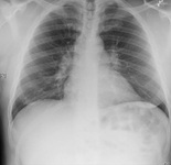
Chest x-ray showing early ill-defined opacities of the right upper lobe above the minor fissure consistent with early changes of aspiration pneumonia
From the personal collection of Dr R. Kanner, University of Utah School of Medicine
See this image in context in the following section/s:

Evaluation of chronic cough
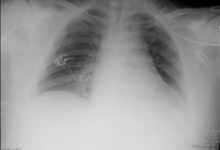
Chest x-ray showing increased opacification of the right perihilar region and superior segment of the right lower and upper lobes consistent with worsening aspiration pneumonia
From the personal collection of Dr R. Kanner, University of Utah School of Medicine
See this image in context in the following section/s:
Evaluation of chronic cough
Chest x-ray showing multiple miliary lung metastases (arrows). The primary tumor was a thyroid carcinoma
E. Dick, Student BMJ. 2001;9:10-12
See this image in context in the following section/s:
Evaluation of chronic cough
Chest x-ray showing a cavitating right hilar carcinoma (arrow)
E. Dick, Student BMJ. 2001;9:10-12
See this image in context in the following section/s:
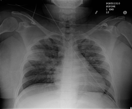
Evaluation of chronic cough
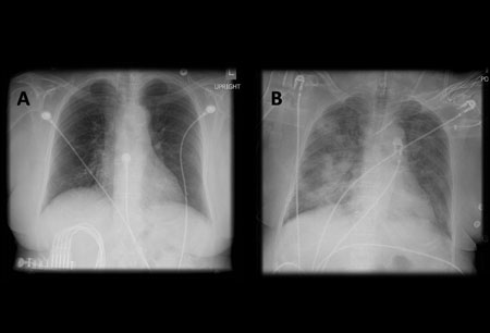
A. Portable upright chest x-ray before aspiration; B. Chest x-ray 1 hour after aspiration, showing bilateral diffuse alveolar infiltrates, worse at the bases on the right side
From the personal collection of Dr S. Murgu and Dr H. Colt, University of California at Irvine Medical Center
See this image in context in the following section/s:
Evaluation of chronic cough
Flow-volume loop (spirogram) in restrictive lung disease (e.g., interstitial pulmonary fibrosis): peak expiratory flow may be normal or low. The shape of the curve is generally normal, but the loop is narrowed and the forced vital capacity is low because of the reduced lung volume.
Created by BMJ Knowledge Centre
See this image in context in the following section/s:

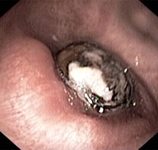
Evaluation of chronic cough
Bronchoscopy image showing a loquat seed completely occluding the bronchus intermedius
From the personal collection of Dr S. Murgu and Dr H. Colt, University of California at Irvine Medical Center
See this image in context in the following section/s:
Evaluation of chronic cough
Chest x-ray showing pulmonary tuberculosis with cavitation
From the personal collection of Dr M. Narita, Department of Pulmonary and Critical Care Medicine, University of Washington
See this image in context in the following section/s:

Evaluation of chronic cough
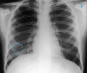
Chest x-ray showing multiple discrete nodules throughout both lungs (one of which is circled) in a patient with miliary tuberculosis
E. Dick, Student BMJ. 2001;9:10-12
See this image in context in the following section/s:
Evaluation of chronic cough
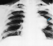
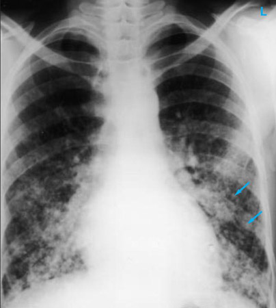
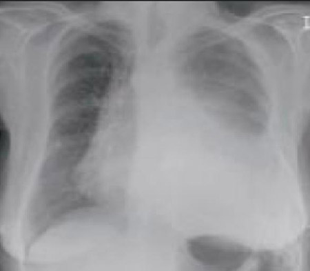
Chest x-ray in a patient with bronchogenic carcinoma showing a left-sided pleural effusion
From: R. Thakkar, Student BMJ. 2001;9:458
See this image in context in the following section/s:
Evaluation of chronic cough
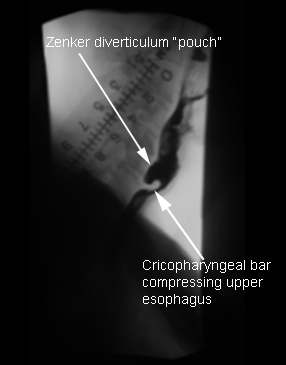
Zenker diverticulum: lateral view with barium esophagram
From the collection of Dr S. Charous, Clinical Professor of Otolaryngology - Head and Neck Surgery, Loyola University Medical Center; used with permission.
See this image in context in the following section/s:

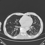
Evaluation of chronic cough
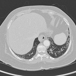
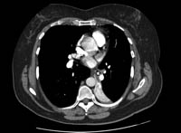
Chest CT with presence of signet ring on left in a patient with bronchiectasis
From the personal collection of Dr S.M. Bhorade, University of Chicago Medical Center
See this image in context in the following section/s:

Evaluation of chronic cough
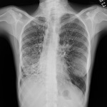
Chest x-ray showing bilateral hilar adenopathy in a patient with sarcoidosis
From the personal collection of Dr M.P. Muthiah, Division of Pulmonary and Critical Care and Sleep Medicine, University of Tennessee
See this image in context in the following section/s:
Evaluation of chronic cough
CT of the chest with intravenous contrast material showing complete left lower lobe collapse with a radiopaque object within the left lower main bronchus surrounded by a halo of air
BMJ Case Reports 2008 (doi:10.1136/bcr.06.2008.0013). Copyright 2008 BMJ Publishing Group Ltd
See this image in context in the following section/s:

Evaluation of chronic cough
Chest CT of a patient with amiodarone pulmonary toxicity, showing asymmetric opacities with a peripheral distribution
From the personal collection of Dr A. Pataka and Professor P. Argyropoulou, Aristotle University, Thessaloniki, Greece
See this image in context in the following section/s:
Evaluation of chronic cough
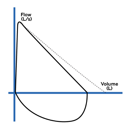
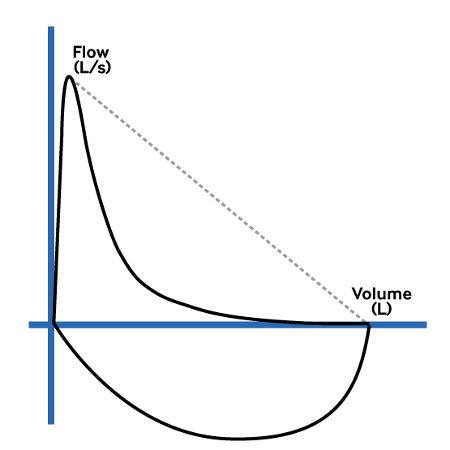
Flow-volume loop (spirogram) in obstructive lung disease, such as asthma or COPD: peak expiratory flow may be normal, but a concave shape is seen following the point of maximal flow due to the low flow rate in relation to lung volume
Created by BMJ Knowledge Centre
See this image in context in the following section/s:

Evaluation of chronic cough
Chest CT showing idiopathic pulmonary fibrosis
From the personal collection of Dr J.C. Munson, Center for Clinical Epidemiology and Biostatistics, University of Pennsylvania School of Medicine
See this image in context in the following section/s:
Evaluation of chronic cough
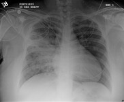
Portable chest x-ray with bibasilar opacities, worse on the right than the left, in a patient with hospital-acquired pneumonia
From the personal collection of Dr F.W. Arnold, Division of Infectious Diseases, Department of Medicine, University of Louisville School of Medicine
See this image in context in the following section/s:

Evaluation of chronic cough
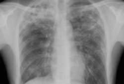
Chest x-ray with dilated and thickened airways in a patient with bronchiectasis
From the personal collection of Dr S.M. Bhorade, University of Chicago Medical Center; used with permission
See this image in context in the following section/s:

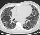
Evaluation of chronic cough
Chest CT with dilated and thickened airways and peripheral tree-in-bud pattern in a patient with bronchiectasis
From the personal collection of Dr S.M. Bhorade, University of Chicago Medical Center; used with permission
See this image in context in the following section/s:

Evaluation of chronic cough
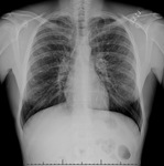
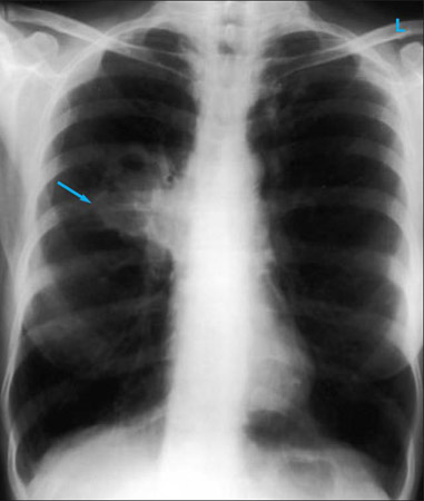
Chest x-ray showing left hilar carcinoma (arrow)
From: E. Dick, Student BMJ. 2000;8:358-360
See this image in context in the following section/s:
Videos
 Peak flow measurement: animated demonstration
How to use a peak flow meter to obtain a peak expiratory flow measurement.
 Venepuncture and phlebotomy: animated demonstration
How to take a venous blood sample from the antecubital fossa using a vacuum needle.
Use of this content is subject to our disclaimer