
Images and videos
Images


Coronavirus disease 2019 (COVID-19)
Virus replication cycle
BMJ. 2020;371:m3862
See this image in context in the following section/s:
Coronavirus disease 2019 (COVID-19)
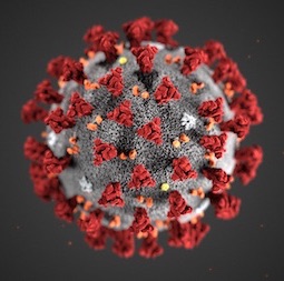
Illustration revealing ultrastructural morphology exhibited by severe acute respiratory syndrome coronavirus 2 (SARS-CoV-2) when viewed with electron microscopically
Centers for Disease Control and Prevention
See this image in context in the following section/s:
Coronavirus disease 2019 (COVID-19)
Number of COVID-19 cases reported weekly by WHO Region, and global deaths, as of 2 February 2025
World Health Organization
See this image in context in the following section/s:
Coronavirus disease 2019 (COVID-19)
Multi-organ complications of COVID-19 and long COVID. The SARS-CoV-2 virus gains entry into the cells of multiple organs via the ACE2 receptor
BMJ. 2021;374:n1648
See this image in context in the following section/s:

Coronavirus disease 2019 (COVID-19)
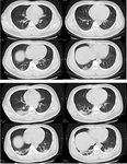
Transverse CT scans from a 32-year-old man, showing ground-glass opacity and consolidation of lower lobe of right lung near the pleura on day 1 after symptom onset (top panel), and bilateral ground-glass opacity and consolidation on day 7 after symptom onset
Xu XW et al. BMJ. 2020;368:m606
See this image in context in the following section/s:
Videos
 Radial artery puncture animated demonstration
How to obtain an arterial blood sample from the radial artery.
Use of this content is subject to our disclaimer