
Approach
Diagnosis is based on characteristic clinical findings, history of exposure in an endemic area, and positive Lyme serology. In an appropriate clinical setting, history and physical exam are sufficient for diagnosis of erythema migrans (EM) and positive serology is not required. Positive Lyme serology requires careful correlation with clinical features to avoid treating patients with a false-positive result.
History and physical exam
Suspect Lyme disease if a tick bite has lasted longer than 36 hours in an endemic area.[25] It is important to be mindful that not all patients recall a tick bite however; if Lyme disease is suspected based on a patient's presentation, conduct a thorough physical exam to look for bites. They can occur anywhere, but are more common on the ankles, behind the knees, and in the groin in adults, and in the head and neck area in children. Check under the hair and behind the ears. Localized lymphadenopathy may occur.[26] Ask about travel, particularly over the past month, to areas where Lyme disease is common, and enquire about recreational or occupational activities that risk exposure to ticks (e.g., outdoor sports, camping, hiking, forestry, and farming). Ticks are common in rural and forested regions with wooded or grassy areas, but can also be found in urban gardens and parks.[26]
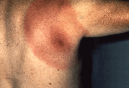
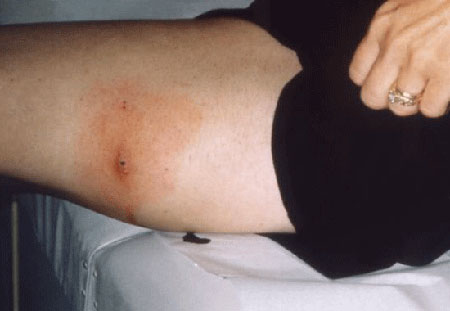
A characteristic rash of erythema migrans in the setting of exposure to ticks in an endemic area is sufficient for clinical diagnosis; in these circumstances, initiate treatment without any further investigation.[27] EM appears in about 50% to 90% of patients with Lyme disease and usually occurs 1 to 2 weeks after a tick bite (range 1 to 36 days).[2] It is often accompanied by symptoms of fatigue, low grade fever, headache, mild stiff neck, arthralgia, or myalgia. These nonspecific viral symptoms may also occur in the absence of rash however, making diagnosis challenging.[28]
Patients may also present with complications of Lyme disease:
Musculoskeletal manifestations include arthralgia and arthritis. Arthritis typically presents with recurrent brief attacks of joint swelling in 1 or more joints, typically involving the knees.
Neurologic manifestations include facial (cranial nerve VII) and other cranial nerve palsies, radiculoneuropathy, lymphocytic (aseptic) meningitis, and encephalitis.
Cardiovascular complications include carditis with atrioventricular block (second- or third-degree) and, less commonly, myopericarditis.
In the US, surveillance of Lyme disease between 2008 and 2015 found that 72.2% of patients had EM, 27.5% had arthritis, 12.5% had neurologic manifestations, and 1.5% had carditis.[6]
Infection with European strains of Borrelia can result in a rare cutaneous manifestation known as borrelial lymphocytoma. This typically presents as a painless bluish-red nodule, papule, or plaque a few centimeters in diameter and is commonly seen on the ear lobe in children and near the nipple area in adults.[1][25]
[Figure caption and citation for the preceding image starts]: Borrelial lymphocytomaGzzz, CC BY-SA 4.0 via Wikimedia Commons; used with permission [Citation ends].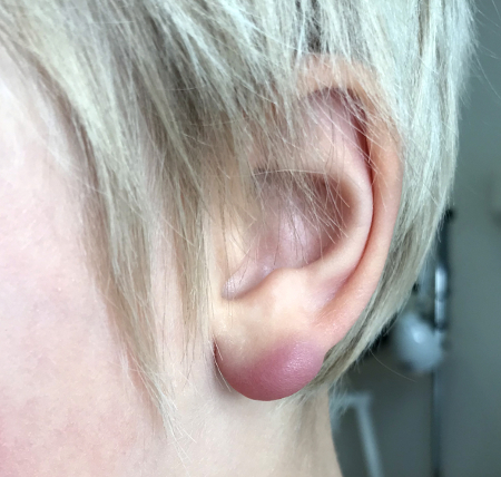
[Figure caption and citation for the preceding image starts]: Acrodermatitis chronica atrophicansNguyen AL et al. Case Reports 2016; 2016: bcr2016216033; used with permission [Citation ends].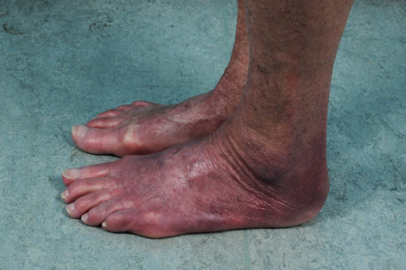
Coinfection with babesiosis or ehrlichiosis (anaplasmosis) may occur. This is because the Ixodes scapularis tick may also transmit Babesia microti and Anaplasma phagocytophila. One study found that in patients with Lyme disease, approximately 2% were infected with B microti and 2% were infected with A phagocytophila.[14] There are often no differentiating symptoms.
[Figure caption and citation for the preceding image starts]: Erythema migransFrom the personal collection of Dr Cristian Speil; used with permission [Citation ends].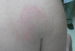 [Figure caption and citation for the preceding image starts]: Erythema migransFrom the personal collection of Dr Cristian Speil; used with permission [Citation ends].
 [Figure caption and citation for the preceding image starts]: Posterior right shoulder region of a patient with Lyme disease showing erythema migransCDC Image Library [Citation ends].
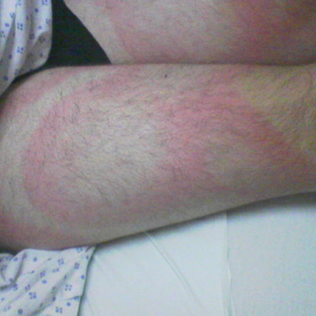
 [Figure caption and citation for the preceding image starts]: Lateral aspect of the left thigh of a patient who’d presented with what was diagnosed as Lyme disease showing the characteristic red, expanding rash (erythema migrans)CDC Image Library [Citation ends].
 [Figure caption and citation for the preceding image starts]: This Lyme disease patient presented with the signs and symptoms indicative of arthritic changes to his right knee due to a Borrelia burgdorferi bacterial infectionCDC Image Library [Citation ends].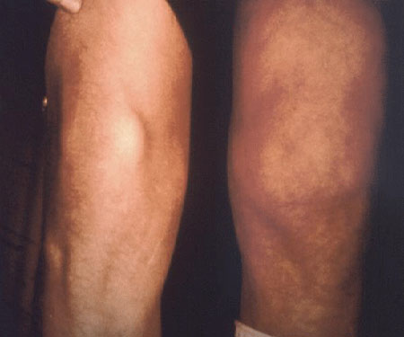
Laboratory testing
In people who live in, or have a history of travel to, an endemic area (with or without a recollection of a tick bite), a diagnosis of Lyme disease can be made by identifying an EM rash. For patients with evidence of disseminated infection (cardiac, musculoskeletal, or neurologic manifestations), serologic testing using commercial assays can aid in diagnosis. The American College of Rheumatology, the American Academy of Pediatrics, and the European Society of Clinical Microbiology and Infectious Diseases study group for Lyme borreliosis recommend against testing individuals with only nonspecific, subjective symptoms.[27][29][30]
Antibody testing
The most readily available diagnostic test. It should be interpreted in combination with clinical symptoms and signs.[1]
Serologic diagnosis includes IgM and IgG antibodies via a 2-tier approach:[5][31][32] CDC: Lyme disease Opens in new window CDC: tickborne diseases Opens in new window
Use a sensitive enzyme immunoassay (EIA) or immunofluorescence assay (IFA) as a first step, and if positive or equivocal, confirm with an immunoblot test (such as a standardized Western blot assay). In the US, some EIAs have been approved by the Food and Drug Administration for serologic diagnosis of Lyme disease and can be used in place of Western blot assays for the second step.[33]
For early Lyme disease (first 4 weeks), test for both IgM and IgG antibodies in the second confirmatory step.
No further testing is needed if specimens are negative by a sensitive EIA or IFA. However, in a patient with suspected early Lyme disease who has a negative EIA, carry out a repeat serologic test during the convalescent phase (paired sera samples) >2 weeks later.
Do not order an immunoblot without first obtaining a positive EIA or IFA screening test result. The immunoblot test is a confirmatory test. Some antigens on the blot react with non-Lyme antibodies, meaning the immunoblot can be falsely interpreted as positive in the absence of a positive initial screening test.[34][35]
People with disseminated or late Lyme disease show a strong IgG response to Borrelia burgdorferi antigens. Because of this, use IgM blots in only the first month of infection.[31] In patients who are persistently symptomatic and are positive only for IgM, carry out a repeat immunoblot after a few weeks. If a repeat test continues to show similar discordant results (IgM+, IgG-), it is likely a false-positive result; consider alternative diagnoses. CDC: Lyme disease Opens in new window False-positive Lyme serology can result from cross-reacting antibodies in autoimmune disorders, infectious mononucleosis, and syphilis.[24] An exposure or infection in the distant past can also result in false-positive serology.[5] These serologic tests have limited sensitivity and specificity.
Patients with neuroborreliosis have over 90% seropositivity. The cerebrospinal fluid (CSF) changes usually show lymphomonocytic leukocytosis with <1000 cells/microliter, and elevated protein and oligoclonal IgG band.[36] In some patients with Lyme disease, antibodies in serum may be passively transferred to CSF; therefore, if neuroborreliosis is suspected, collect CSF and serum on the same day and dilute to match total IgG concentration. A CSF/serum IgG ratio of >1.0 indicates active intrathecal antibody production; however, there is no agreement on a diagnostic cut-off threshold.[25][32][34] A CSF/serum antibody index ratio of >1.3 has been used as a threshold for supporting diagnosis of neuroborreliosis. Small studies have shown sensitivity of only 53% and specificity of 100% with a cut-off of 1.3, and sensitivity of 87% and specificity of 93% using 0.9.[37]
Although a vaccine against Lyme disease is no longer available, previous administration of OspA vaccine (LYMErix) may affect test results, resulting in false-positive ELISA tests and a positive band (or multiple bands) on Western blot tests for IgG. If coinfection with B microti or A phagocytophila is suspected, serology is recommended to confirm diagnosis of babesiosis or ehrlichiosis. Peripheral blood smear and PCR are also recommended.
Microbiological culture
Culture of infected tissue is a more direct test than serologic diagnosis, but it is not recommended routinely because it is invasive, costly, and more difficult to carry out.[25] Culture requires special media, takes a long time (up to 8 weeks or longer), and is generally not available. A positive culture result is likely with skin biopsy specimens taken from erythema migrans lesions, but is less likely with serum and cerebrospinal fluid samples.[38] There are only anecdotal reports about joint fluid samples yielding positive results.
Polymerase chain reaction (PCR)
PCR is generally superior to culture for specimens such as joint fluid, skin biopsy, and other solid tissue specimens, but it is not widely available and requires referral to specialists for biopsy.[25][37] Sensitivity of PCR is modest (50% to 70%) for joint fluid and skin specimens, but poor for blood and CSF (10% to 30%).[5][32][34] Where available, PCR assay of skin and other tissue biopsy specimens can be used as an adjunctive test in select patients to confirm the diagnosis when serology is positive but clinical presentation is atypical.
ECG
An ECG is indicated only in patients with signs and symptoms of cardiac disease, such as shortness of breath, edema, chest pain, palpitations, lightheadedness, or syncope.[25] ECG findings typically include acute onset of varying degrees of intermittent atrioventricular block with rapidly fluctuating complete heart block. Atrial and ventricular arrhythmias may occur. Myopericarditis occurs rarely.
Use of this content is subject to our disclaimer