Резюме
Определение
Кольцевидная гранулема (КГ) является редким доброкачественным образованием неизвестной этиологии.[1] Поражения обычно описываются как кожные папулы, которые сливаются в форме колец и могут иметь телесный, розовый или фиолетовый цвет; часто возникают на тыльной стороне ладони, лодыжках, коленях и локтях.[Figure caption and citation for the preceding image starts]: Классическая локализованная кольцевидная гранулемаИз коллекции Сусмита Мукхерджи (Susmita Mukherjee), бакалавра наук, бакалавра медицины и бакалавра хирургии, магистра, члена Королевского колледжа врачей; используется с разрешения [Citation ends].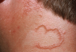 Существует ряд клинических форм.[2] Данные гистологического исследования являются отличительными чертами. Большинство случаев заканчиваются самоизлечением.[3]
Анамнез и осмотр
Ключевые диагностические факторы
- бессимптомные сгруппированные аннулярные дермальные папулы розового или телесного цвета
Другие диагностические факторы
- телесного цвета, розовый или коричневый macules или небольшие папулы
- узлы в мягких тканях
- перфорирующие папулы, корочки или язвенные поражения
- эритематозные пятна
Факторы риска
- сахарный диабет
- Злокачественное заболевание крови
- опоясывающий лишай
- ВИЧ
- гепатит
- гиперлипидемия
- заболевания щитовидной железы
- лекарственные препараты
Диагностические исследования
Исследования, которые показаны в первую очередь
- клинический диагноз
Исследования, проведение которых нужно рассмотреть
- биопсия кожи
- уровень сахара в крови натощак
- исследование функции щитовидной железы
- скрининг липидов
- скрининг гепатита
- исследование на ВИЧ
Алгоритм лечения
локализованная форма (классическая кольцевая сыпь)
генерализованная форма (распространенная макулярная сыпь)
перфорирующая форма (корочки или язвенные поражения)
подкожная форма (мягкий узелок)
пятнистая форма (эритематозные бляшки)
Составители
Авторы
Misha Rosenbach, MD
Associate Professor
Dermatology and Internal Medicine
University of Pennsylvania
Philadelphia
PA
Раскрытие информации
MR has received consulting fees from Merck, aTyr Pharma, and Processa Pharma, not related to granuloma annulare. He has received grant support from Processa Pharma, not related to granuloma annulare. MR has received salary support from the AMA/JAMA for his role at JAMA Dermatology as Deputy Editor. MR is an author of references cited in this topic.
Выражение благодарностей
Dr Misha Rosenbach would like to gratefully acknowledge Dr Susmita Mukherjee, a previous contributor to this topic. SM declares that she has no competing interests.
Рецензенты
Aisha Sethi, MD
Assistant Professor of Medicine
Associate Residency Program Director
University of Chicago
Chicago
IL
Раскрытие информации
AS declares that she has no competing interests.
Robert Herd, MSc, MD, FRCP
Consultant Dermatologist
Department of Dermatology
Nuffield Hospital
Glasgow
UK
Раскрытие информации
RH declares that he has no competing interests.
Brenda L. Pellicane, MD
Dermatologist
Wayne State University School of Medicine
Department of Dermatology
Detroit
MI
Раскрытие информации
BLP declares that she has no competing interests.
Peer reviewer acknowledgements
BMJ Best Practice topics are updated on a rolling basis in line with developments in evidence and guidance. The peer reviewers listed here have reviewed the content at least once during the history of the topic.
Disclosures
Peer reviewer affiliations and disclosures pertain to the time of the review.
Список литературы
Основные статьи
Piette EW, Rosenbach M. Granuloma annulare: clinical and histologic variants, epidemiology, and genetics. J Am Acad Dermatol. 2016 Sep;75(3):457-65. Аннотация
Thornsberry LA, English JC 3rd. Etiology, diagnosis, and therapeutic management of granuloma annulare: an update. Am J Clin Dermatol. 2013 Aug;14(4):279-90. Аннотация
Piette EW, Rosenbach M. Granuloma annulare: pathogenesis, disease associations and triggers, and therapeutic options. J Am Acad Dermatol. 2016 Sep;75(3):467-79. Аннотация
Статьи, указанные как источники
A full list of sources referenced in this topic is available to users with access to all of BMJ Best Practice.
Отличия
- Кольцевидный плоский лишай
- Дерматофитные инфекции
- Грибковый микоз
Больше Отличия Войдите в учетную запись или оформите подписку, чтобы получить полноценный доступ к BMJ Best Practice
Использование этого контента попадает под действие нашего заявления об отказе от ответственности