Resumo
Definição
História e exame físico
Principais fatores diagnósticos
- presença de fatores de risco
- perda da visão súbita e indolor
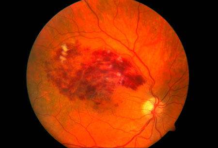
- edema da cabeça do nervo óptico
- hemorragia intrarretiniana
- tortuosidade e dilatação venosas
- formação de vasos colaterais
Outros fatores diagnósticos
- idade >65 anos
- neovascularização
- hemorragia do vítreo
- edema macular
- moscas volantes
- olho dolorido e vermelho
- acuidade visual
- defeito pupilar aferente relativo
- pressão intraocular elevada
Fatores de risco
- aterosclerose
- hipertensão sistêmica
- diabetes mellitus
- hiperlipidemia
- história de tabagismo
- doença cardiovascular
- glaucoma
- índice de massa corporal aumentado aos 20 anos de idade
- aumento de alfa-2-globulina sérica
- comprimento axial curto
- idade >65 anos
- resistência à proteína C ativada
- fator V de Leiden
- estado hipercoagulável
- deficiência de proteína S
- deficiência de antitrombina III
- hiper-homocisteinemia
- vasculite
Investigações diagnósticas
Primeiras investigações a serem solicitadas
- angiografia com fluoresceína para confirmação do diagnóstico
Investigações a serem consideradas
- angiografia com fluoresceína para avaliação de complicações
- tomografia de coerência óptica
- eletrorretinografia
Algoritmo de tratamento
OVRC: não complicada
oclusão da veia retiniana central (OVRC) com edema macular
OVRC com neovascularização
ORVR ou OVHR: não complicada
oclusão de ramo da veia retiniana (ORVR) ou oclusão da veia hemirretiniana (OVHR) com edema macular
oclusão de ramo da veia retiniana (ORVR) ou oclusão da veia hemirretiniana (OVHR) com neovascularização
Colaboradores
Autores
Aziz A. Khanifar, MD

Vitreoretinal Surgeon
The Retina Group of Washington
Silver Spring
MD
Declarações
AAK has been a speaker for Genentech and Regeneron.
Agradecimentos
Dr Aziz A. Khanifar would like to gratefully acknowledge Dr Sharon Fekrat, a previous contributor to this topic.
Declarações
SF declares that she has no competing interests.
Revisores
Stephen G. Schwartz, MD
Assistant Professor of Clinical Ophthalmology
Bascom Palmer Eye Institute
University of Miami Miller School of Medicine
Naples
FL
Declarações
SGS has received research funding from Alcon, Genentech, and Novartis; holds equity in Pfizer; and is co-holder of a patent pending entitled "Molecular targets for modulating intraocular pressure and differentiation of steroid responders from non-responders".
Tim Murray, MD
Professor of Ophthalmology
Bascom Palmer Eye Institute
University of Miami Miller School of Medicine
Naples
FL
Declarações
TM declares that he has no competing interests.
Alexander Spratt, MBBCh, MRCOphth
Specialist Registrar
Moorfields Eye Hospital
Visiting Lecturer
City University
London
UK
Declarações
AS has received research funding from Allergan.
Créditos aos pareceristas
Os tópicos do BMJ Best Practice são constantemente atualizados, seguindo os desenvolvimentos das evidências e das diretrizes. Os pareceristas aqui listados revisaram o conteúdo pelo menos uma vez durante a história do tópico.
Declarações
As afiliações e declarações dos pareceristas referem--se ao momento da revisão.
Referências
Principais artigos
Romano F, Lamanna F, Gabrielle PH, et al. Update on retinal vein occlusion. Asia Pac J Ophthalmol (Phila). 2023 Mar-Apr;12(2):196-210.Texto completo Resumo
The Royal College of Opthalmologists. Retinal vein occlusion guidelines. Feb 2022 [internet publication].Texto completo
Kovach JL, Bailey ST, Kim SJ, et al. Retinal vein occlusions preferred practice pattern®. Ophthalmology. 2025 Apr;132(4):P303-43.Texto completo
Nicholson L, Talks SJ, Amoaku W, et al. Retinal vein occlusion (RVO) guideline: executive summary. Eye (Lond). 2022 May;36(5):909-12.Texto completo Resumo
Shalchi Z, Mahroo O, Bunce C, et al. Anti-vascular endothelial growth factor for macular oedema secondary to branch retinal vein occlusion. Cochrane Database Syst Rev. 2020 Jul 7;7:CD009510.Texto completo Resumo
Artigos de referência
Uma lista completa das fontes referenciadas neste tópico está disponível para os usuários com acesso total ao BMJ Best Practice.
Diagnósticos diferenciais
- Neuropatia óptica isquêmica
- Descolamento da retina (DR)
- Neovascularização da coroide (NVC)
Mais Diagnósticos diferenciaisDiretrizes
- Retinal vein occlusions preferred practices pattern
- Retina summary benchmarks - 2024
Mais DiretrizesFolhetos informativos para os pacientes
Diabetes: o que posso fazer para me manter saudável?
Pressão alta: o que é?
Mais Folhetos informativos para os pacientes Conectar-se ou assinar para acessar todo o BMJ Best Practice
O uso deste conteúdo está sujeito ao nosso aviso legal