Resumo
Definição
História e exame físico
Principais fatores diagnósticos
- presença de fatores de risco
- perda ou deterioração da visão central
- flashes de luz
- perda do campo visual periférico
Outros fatores diagnósticos
- moscas volantes
Fatores de risco
- descolamento do vítreo posterior
- idade mais avançada
- miopia alta
- cirurgia oftálmica prévia
- trauma
- tumor intraocular
- hemorragia do vítreo
- olho contralateral afetado
- diabetes mellitus
- retinopatia da prematuridade
- história familiar de descolamento de retina não traumático em familiar próximo
- inflamação/infecção ocular
- degeneração retiniana periférica
- anormalidade anatômica
- degeneração macular relacionada à idade
- uso de inibidor da fosfodiesterase-5 em homens
- causas genéticas e vasculares na infância
- tumores na infância
Investigações diagnósticas
Primeiras investigações a serem solicitadas
- teste da acuidade visual
- exame com lâmpada de fenda
- oftalmoscopia indireta
Investigações a serem consideradas
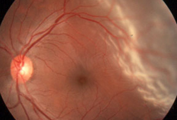
- fotografia colorida de campo amplo
- tomografia de coerência óptica (olho afetado)
- Ultrassonografia B scan (olho afetado)
- tomografia computadorizada (TC)/ressonância nuclear magnética (RNM) da órbita
Algoritmo de tratamento
descolamento do vítreo posterior sem ruptura
ruptura da retina sem descolamento
descolamento de retina (DR) regmatogênico
descolamento de retina (DR) tracional
descolamento de retina (DR) exsudativo
descolamento de retina (DR) hemorrágico
Colaboradores
Autores
Ferenc Kuhn, MD, PhD

Director of Clinical Research
Helen Keller Foundation for Research and Education
Associate Professor of Ophthalmology
University of Alabama at Birmingham
Birmingham
AL
Consultant and Chief Vitreoretinal Surgeon
Department of Ophthalmology
University of Pécs Medical School
Pécs
Hungary
Declarações
FK declares that he has no competing interests.
Agradecimentos
Dr Ferenc Kuhn would like to gratefully acknowledge Dr Robert Morris, a previous contributor to this topic.
Declarações
RM declares that he has no competing interests.
Revisores
David Steel, MBBS, FRCOphth
Consultant Ophthalmologist
Sunderland Eye Infirmary
Sunderland
UK
Declarações
DS declares that he has no competing interests.
Michael W. Stewart, MD
Professor and Chairman of Ophthalmology
Mayo Clinic
Jacksonville
FL
Declarações
MWS declares that he has no competing interests.
Ron Adelman, MD, MPH, FACS
Associate Professor of Ophthalmology
Yale University School of Medicine
New Haven
CT
Declarações
RA declares that he has no competing interests.
Scott Fraser, MD, FRCS (Ed), FRCOphth
Consultant Ophthalmologist
Sunderland Eye Infirmary
Sunderland
UK
Declarações
SF declares that he has no competing interests.
Créditos aos pareceristas
Os tópicos do BMJ Best Practice são constantemente atualizados, seguindo os desenvolvimentos das evidências e das diretrizes. Os pareceristas aqui listados revisaram o conteúdo pelo menos uma vez durante a história do tópico.
Declarações
As afiliações e declarações dos pareceristas referem--se ao momento da revisão.
Referências
Principais artigos
Kim SJ, Bailey ST, Kovach JL, et al. Posterior vitreous detachment, retinal breaks, and lattice degeneration preferred practice pattern®. Ophthalmology. 2025 Apr;132(4):P163-96.Texto completo
American Academy of Ophthalmology. Referral of persons with possible eye diseases or injury - 2014. Apr 2014 [internet publication].Texto completo
Hikichi T, Trempe CL. Relationship between floaters, light flashes, or both, and complications of posterior vitreous detachment. Am J Ophthalmol. 1994 May 15;117(5):593-8. Resumo
Znaor L, Medic A, Binder S, et al. Pars plana vitrectomy versus scleral buckling for repairing simple rhegmatogenous retinal detachments. Cochrane Database Syst Rev. 2019 Mar 8;(3):CD009562.Texto completo Resumo
Sena DF, Kilian R, Liu SH, et al. Pneumatic retinopexy versus scleral buckle for repairing simple rhegmatogenous retinal detachments. Cochrane Database Syst Rev. 2021 Nov 11;(11):CD008350.Texto completo Resumo
Artigos de referência
Uma lista completa das fontes referenciadas neste tópico está disponível para os usuários com acesso total ao BMJ Best Practice.
Diagnósticos diferenciais
- Tração vitreomacular
- Retinosquise
- Retinopatia diabética
Mais Diagnósticos diferenciaisDiretrizes
- Comprehensive adult medical eye evaluation preferred practice pattern
- Retina summary benchmarks - 2025
Mais DiretrizesFolhetos informativos para os pacientes
Degeneração macular
Mais Folhetos informativos para os pacientes Conectar-se ou assinar para acessar todo o BMJ Best Practice
O uso deste conteúdo está sujeito ao nosso aviso legal