Summary
Definition
History and exam
Key diagnostic factors
- presença de fatores de risco
- perda de campo visual
Other diagnostic factors
- cefaleia
- obscurecimentos visuais transitórios
- zumbido síncrono ao pulso
- fotofobia
- dor retrobulbar
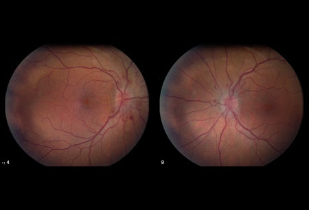
- edema do disco óptico
- acuidade visual reduzida
- distúrbios de motilidade ocular
- defeito pupilar aferente relativo
Risk factors
- sexo feminino
- obesidade e ganho de peso
- uso de determinados medicamentos
- doenças causais associadas
- apneia do sono
- história familiar
Diagnostic investigations
1st investigations to order
- testes de campo visual (perimetria)
- fundoscopia dilatada
- acuidade visual
- ressonância nuclear magnética (RNM) cranioencefálica com ou sem contraste
- punção lombar nos segmentos L3/L4 da espinha
Investigations to consider
- venografia por ressonância magnética da cabeça
- tomografia de coerência óptica
Treatment algorithm
todos os pacientes
Contributors
Authors
Michael Wall, MD
Professor
Department of Neurology and Department of Ophthalmology & Visual Sciences
University of Iowa Hospitals & Clinics and Iowa City VA Health Care System
Iowa City
IA
Disclosures
MW is an author of a number of references cited in this topic.
Mansoor Mughal, MD
Retina Fellow
Rutgers University
Robert Wood Johnson University Hospital
New Brunswick
NJ
Disclosures
MM declares that he has no competing interests.
Peer reviewers
Paul W. Brazis, MD
Consultant in Neurology and Neuro-Ophthalmology
Mayo Clinic Florida
Jacksonville
FL
Disclosures
PWB declares that he has no competing interests.
Tim D. Matthews, MBBS
Consultant Neuro-ophthalmologist
Birmingham Neuro-ophthalmology Unit
University Hospital Birmingham
Birmingham
UK
Disclosures
TDM declares that he has no competing interests.
Peer reviewer acknowledgements
BMJ Best Practice topics are updated on a rolling basis in line with developments in evidence and guidance. The peer reviewers listed here have reviewed the content at least once during the history of the topic.
Disclosures
Peer reviewer affiliations and disclosures pertain to the time of the review.
References
Key articles
Wall M. Idiopathic intracranial hypertension. Neurol Clin. 2010 Aug;28(3):593-617.Full text Abstract
Johnson LN, Krohel GB, Madsen RW, et al. The role of weight loss and acetazolamide in the treatment of idiopathic intracranial hypertension (pseudotumor cerebri). Ophthalmology. 1998 Dec;105(12):2313-7. Abstract
Hayreh SS. Pathogenesis of oedema of the optic disc (papilloedema): a preliminary report. Br J Ophthalmol. 1964 Oct;48:522-43.Full text Abstract
Smith JL. Whence pseudotumor cerebri? J Clin Neuroophthalmol. 1985 Mar;5(1):55-6. Abstract
Frisen L. Swelling of the optic nerve head: a staging scheme. J Neurol Neurosurg Psychiatry. 1982 Jan;45(1):13-8.Full text Abstract
NORDIC Idiopathic Intracranial Hypertension Study Group Writing Committee; Wall M, McDermott MP, Kieburtz KD, et al. Effect of acetazolamide on visual function in patients with idiopathic intracranial hypertension and mild visual loss: the idiopathic intracranial hypertension treatment trial. JAMA. 2014 Apr 23-30;311(16):1641-51.Full text Abstract
Sinclair AJ, Burdon MA, Nightingale PG, et al. Low energy diet and intracranial pressure in women with idiopathic intracranial hypertension: prospective cohort study. BMJ. 2010 Jul 7;341:c2701.Full text Abstract
Hoffmann J, Mollan SP, Paemeleire K, et al. European Headache Federation guideline on idiopathic intracranial hypertension. J Headache Pain. 2018 Oct 8;19(1):93.Full text Abstract
Reference articles
A full list of sources referenced in this topic is available to users with access to all of BMJ Best Practice.
Differentials
- Anomalias estruturais intracranianas
More DifferentialsGuidelines
- European Headache Federation guideline on idiopathic intracranial hypertension
More GuidelinesPatient information
Zumbido
Obesidade - medicamentos e cirurgia
More Patient information Log in or subscribe to access all of BMJ Best Practice
Use of this content is subject to our disclaimer