Summary
Definition
History and exam
Key diagnostic factors
- hyphema
- ecchymosis
- severe eye pain
- blurred vision
- corneal abrasions
- corneal edema
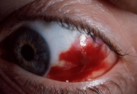
- subconjunctival hemorrhages
- corneal and conjunctival lacerations
- punctate epithelial erosions
- loss of sight
Other diagnostic factors
- excessive lacrimation
- conjunctival chemosis
- conjunctival hyperemia
- corneal epithelial defect/abrasion
- open globe injury
- eyelid burns
- photophobia
- diplopia
- miosis
- corneal stromal clouding
- iridodialysis
- conjunctival foreign body
- corneal foreign body
- Descemet membrane tears
- corneoscleral lacerations
- persistent headache
- loss of consciousness
- blood or clear fluid from ears or nose
- inability to move eye(s)
Risk factors
- age 18-45 years
- male sex
- no protective eyewear
- workplace injuries
- falls
- fireworks
- exposure to ultraviolet light
- previous eye surgery
- alcohol-based hand sanitizers
Diagnostic tests
1st tests to order
- CT scan of orbit
- CT scan of head
- MRI scan of head
Tests to consider
- plain x-ray
- B-scan ultrasonography
- ultrasound biomicroscopy
- optical coherence tomography
- fluorescein angiography
- fundus autofluorescence
- urine drug screen
- sickle cell trait screen
Treatment algorithm
superficial injuries without a foreign body
hyphema
corneal abrasion
open globe injury
recurrent corneal erosions or poor healing
Contributors
Authors
Fasika Woreta, MD, MPH
Associate Professor of Ophthalmology
Residency Program Director
Director, Eye Trauma Center
Wilmer Eye Institute
Johns Hopkins University School of Medicine
Baltimore
MD
Disclosures
FW declares that she has no competing interests.
Acknowledgements
Dr Fasika Woreta wishes to gratefully acknowledge Dr Ron Adelman and Dr Elena Raluca Raducu, previous contributors to this topic.
Disclosures
ERR declares that she has no competing interests.
Peer reviewers
Yewlin E. Chee, MD
Assistant Professor of Ophthalmology
University of Washington
Seattle
WA
გაფრთხილება:
YE declares that she has no competing interests.
Andrew W. Eller, MD
Professor of Ophthalmology
University of Pittsburgh School of Medicine
Pittsburgh
PA
გაფრთხილება:
AE declares that he has no competing interests.
Saloni Kapoor, MD
Clinical Assistant Professor of Medicine
UPMC Mercy Hospital
Pittsburgh
PA
გაფრთხილება:
SK declares that she has no competing interests.
Seanna Grob, MD, MAS
Assistant Professor of Ophthalmology
Oculoplastic Surgeon
University of California San Francisco
San Francisco
CA
გაფრთხილება:
SG has written a book on ocular trauma.
რეცენზენტების განცხადებები
BMJ Best Practice-ის თემების განახლება სხვადასხვა პერიოდულობით ხდება მტკიცებულებებისა და რეკომენდაციების განვითარების შესაბამისად. ქვემოთ ჩამოთვლილმა რეცენზენტებმა თემის არსებობის მანძილზე კონტენტს ერთხელ მაინც გადახედეს.
გაფრთხილება
რეცენზენტების აფილიაციები და გაფრთხილებები მოცემულია გადახედვის მომენტისთვის.
წყაროები
ძირითადი სტატიები
Kuhn F, Mester V, Berta A, et al. Epidemiology of severe eye injuries: the United States Eye Injury Registry (USEIR) and the Hungarian Eye Injury Registry (HEIR) [in German]. Ophthalmologe. 1998 May;95(5):332-43. აბსტრაქტი
Kuhn F, Morris R, Witherspoon CD. Birmingham Eye Trauma Terminology (BETT): terminology and classification of mechanical eye injuries. Ophthalmol Clin North Am. 2002 Jun;15(2):139-43. აბსტრაქტი
MacEwen CJ. Ocular injuries. J R Coll Surg Edinb. 1999 Oct;44(5):317-23. აბსტრაქტი
American Academy of Ophthalmology. Policy statement: referral of persons with possible eye diseases or injury. Apr 2014 [internet publication].სრული ტექსტი
გამოყენებული სტატიები
ამ თემაში მოხსენიებული წყაროების სრული სია ხელმისაწვდომია მომხმარებლებისთვის, რომლებსაც აქვთ წვდომა BMJ Best Practice-ის ყველა ნაწილზე.

გაიდლაინები
- Pediatric eye evaluations preferred practice pattern
- RCH clinical practice guidelines: acute eye injury
მეტი გაიდლაინებიშედით სისტემაში ან გამოიწერეთ BMJ Best Practice
ამ მასალის გამოყენება ექვემდებარება ჩვენს განცხადებას