Summary
Monoclonal gammopathies represent a wide spectrum of related diseases.[1][2] The common denominator is the presence of a monoclonal protein in the serum or urine, which can be in the form of intact immunoglobulin, immunoglobulin fragments, and/or free light chains. This will be accompanied by the presence of monoclonal plasma cells in the bone marrow (bone plasmacytoma), in soft tissue (extramedullary plasmacytoma), or in the peripheral circulation (typically in more advanced disease stages).
Plasma cells and monoclonal proteins
Plasma cells are terminally differentiated (specialized cells that typically do not proliferate) effector cells of B-cell lineage.[3][4][5][6] They are the primary mediators of humoral immunity, secreting antigen-specific immunoglobulins. Abnormalities of plasma cells are responsible for a variety of autoimmune diseases and plasma cell neoplasms. Clonal evolution of one or more plasma cells sets the stage for development of the monoclonal gammopathies.[3][4][5][6]
Plasma cells normally secrete an intact immunoglobulin that is made up of 2 identical light chains and 2 identical heavy chains. There are 5 major classes of heavy chains, which correspond to the major classes of immunoglobulins: mu (IgM), delta (IgD), gamma (IgG), alpha (IgA), and epsilon (IgE). In each of the immunoglobulin molecules, the heavy chains are bound to one of the 2 light chains (kappa or lambda), but not both. The heavy chains, which have 4 or 5 domains, and the light chains, which have 2 domains, are covalently bonded to each other through disulfide bonds.[Figure caption and citation for the preceding image starts]: Immunoglobulin structureFrom the personal collection of Dr Kumar [Citation ends].
Monoclonal proteins are abnormal, immunologically homogeneous immunoglobulins or parts of immunoglobulins, produced by a single clone of plasma cells. They may be the result of an underlying lymphoid malignancy, be part of a clonal expansion of plasma cells causing no symptoms (e.g., monoclonal gammopathy of unknown significance), or lead to life-threatening complications (e.g., primary amyloidosis).[7]
Evaluation of monoclonal proteins
Protein electrophoresis is performed to detect and identify monoclonal proteins in the serum and urine.[8][9] Quantitation of immunoglobulins is achieved by nephelometry. Immunoglobulin free light chains in serum are assessed using antibodies specific for the light chain part of the immunoglobulins.
Mass spectrometry is increasingly used for the detection and quantitation of monoclonal proteins in serum and urine.[10][11]
Clonal expansion of plasma cells
Clonal expansion of plasma cells is the underlying abnormality among the monoclonal gammopathies. These cells may be found in the bone marrow, soft tissue, or peripheral circulation. They are typically demonstrated on bone marrow exams, where presence of clonal plasma cells may or may not be accompanied by an absolute increase in the plasma cell proportion. Whereas plasma cells are identified by their surface staining for CD138 (syndecan) on immunohistochemistry, demonstration of clonality depends on light chain restriction, and an excess of kappa- or lambda-expressing plasma cells resulting in a skewing of the normal kappa to lambda ratio.[12] The skewed ratio can be demonstrated by immunohistochemistry, flow cytometry, or polymerase chain reaction (PCR) performed on bone marrow aspirate or biopsy.
Flow cytometry techniques can be used for the detection of small clonal plasma cell numbers (typical of most monoclonal gammopathies) in bone marrow aspirate, and for immunophenotypic characterization of abnormal plasma cells.[13][14][15][16][17]
[Figure caption and citation for the preceding image starts]: Monoclonal plasma cellsFrom the personal collection of Dr Kumar [Citation ends].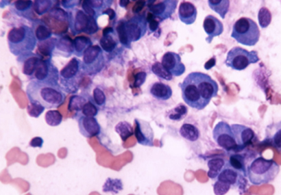
Multiple myeloma and plasma cell leukemia are associated with large numbers of malignant plasma cells in the blood and bone marrow.[18]
Epidemiology
Monoclonal gammopathy of undetermined significance (MGUS) is the most common monoclonal gammopathy and is found in approximately 2% to 3% of the white population age 50 years and older.[19][20][21]
The prevalence increases with age and is generally higher in males compared with females, and in people over 70 years of age.[22][23] Prevalence rates vary according to geographic and racial factors, with lower rates in Asia compared with those in Europe and North and South America, and higher prevalence rates among black people compared with white people.[24]
A total monoclonal gammopathy prevalence of 43% has been reported in a US cohort (at high risk for multiple myeloma; median age 56 years) screened by quantitative mass spectrometry.[25]
Increased prevalence has been observed among first-degree relatives of patients with monoclonal gammopathies.[26][27]
Associated conditions
Other conditions where a monoclonal protein may be detected in the serum and/or urine include:[28][29][30][31][32][33]
lymphoproliferative disease, where clonal B lineage cells secrete a monoclonal protein (e.g., chronic lymphocytic leukemia, non-Hodgkin lymphoma, post-transplant monoclonal gammopathies)
infectious or inflammatory conditions associated with a transient development of several clones of reactive B cell/plasma cell populations (e.g., systemic lupus erythematosus, rheumatoid arthritis, psoriatic arthritis, Sjogren syndrome).
Chronic viral infection may predispose to an increased risk for monoclonal gammopathy (e.g., hepatitis C virus infection, HIV infection).[34][35][36]
Differentials
Common
- Monoclonal gammopathy of undetermined significance
- Chronic lymphocytic leukemia
- Non-Hodgkin lymphoma
Uncommon
- Light chain deposition disease
- Post-transplant monoclonal gammopathies
- Multiple myeloma (MM)
- Plasma cell leukemia
- Waldenström macroglobulinemia (WM)
- Solitary plasmacytoma
- POEMS syndrome (polyneuropathy, organomegaly, endocrinopathy, M protein, and skin changes)
- Hepatitis C
- HIV infection
- Primary amyloidosis
- Cryoglobulinemia
- Systemic lupus erythematosus (SLE)
- Rheumatoid arthritis
- Psoriatic arthritis
- Sjogren syndrome
Contributors
Authors
Shaji Kumar, MD
Mark and Judy Mullins Professor of Hematological Malignancies
Division of Hematology
Mayo Clinic
Rochester
MN
Disclosures
SK has consulted for the following organizations with no personal payment: AbbVie, Amgen, ArcellX, Beigene, BMS, Carsgen, GSK, Janssen, K36, Moderna, Pfizer, Regeneron, Roche-Genentech, Sanofi, Takeda, (with personal payments) CVS Caremark, BD Biosciences. SK has received research funding for clinical trials for his institution from: AbbVie, Amgen, Astra Zeneca, BMS, Carsgen, GSK, Gracell Bio, Janssen, Oricell, Roche-Genentech, Sanofi, Takeda, Telogenomics.
Peer reviewers
Antonio Palumbo, MD
Associate Professor
University of Turin
Chief
Myeloma Unit
Ospedale Molinette
Turin
Italy
Disclosures
AP declares that he has no competing interests.
John R. Wingard, MD
Price Eminent Scholar and Professor of Medicine Director
Bone Marrow Transplant Program
Division of Hematology and Oncology
University of Florida College of Medicine
Gainesville
FL
Disclosures
JRW declares that he has no competing interests.
John Densmore, MD, PhD
Associate Professor of Clinical Medicine
Department of Medicine
Division of Hematology/Oncology
University of Virginia
Charlottesville
VA
Disclosures
JD declares that he has no competing interests.
Peer reviewer acknowledgements
BMJ Best Practice topics are updated on a rolling basis in line with developments in evidence and guidance. The peer reviewers listed here have reviewed the content at least once during the history of the topic.
Disclosures
Peer reviewer affiliations and disclosures pertain to the time of the review.
References
Key articles
Keren DF, Bocsi G, Billman BL, et al. Laboratory detection and initial diagnosis of monoclonal gammopathies. Arch Pathol Lab Med. 2022 May 1;146(5):575-90.Full text Abstract
Myeloma UK. Myeloma, MGUS and related conditions: a guide for GPs. Apr 2025 [internet publication].Full text
Stern S, Chaudhuri S, Drayson M, et al. Investigation and management of the monoclonal gammopathy of undetermined significance: a British Society for Haematology Good Practice Paper. Br J Haematol. 2023 Aug;202(4):734-44.Full text Abstract
Liu Y, Parks AL. Diagnosis and management of monoclonal gammopathy of undetermined significance: a review. JAMA Intern Med. 2025 Apr 1;185(4):450-6.Full text Abstract
Reference articles
A full list of sources referenced in this topic is available to users with access to all of BMJ Best Practice.
Patient information
Monoclonal gammopathy of undetermined significance
Non-Hodgkin lymphoma
More Patient information Log in or subscribe to access all of BMJ Best Practice
Use of this content is subject to our disclaimer