Summary
Definition
History and exam
Key diagnostic factors
- pruritus
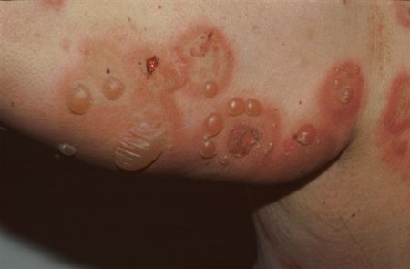
- tense blisters on normal or erythematous skin
Other diagnostic factors
- erythematous or urticarial plaques
- oral lesions
Risk factors
- age 60 to 90 years
- major histocompatibility complex (MHC) class II allele (DQB1*0301)
- neurologic conditions
- use of certain drugs
- male sex
Diagnostic tests
1st tests to order
- skin biopsy for histopathologic evaluation with light microscopy
- skin biopsy for direct immunofluorescence testing
Tests to consider
- ELISA test
- indirect immunofluorescence test on serum
Treatment algorithm
localized lesions in children or adults
widespread lesions in adults
widespread lesions in children
Contributors
Authors
Kyle T. Amber, MD, FAAD
Associate Professor of Dermatology
Vice Chair of Research
Director, Dermatology Infusion Center
Rush University
Chicago, IL
Disclosures
KTA has received research support from Argenx, AstraZeneca, Sanofi, Eli Lilly, and KabaFusion. He also consults for Argenx, AstraZeneca, KabaFusion, Caribou, and Akari and has been a speaker for Argenx and KabaFusion.
Acknowledgements
Dr Kyle T. Amber would like to gratefully acknowledge Dr Vesna Petronic-Rosic, the previous contributor to this topic. VPR declares that she has no competing interests.
Peer reviewers
Lawrence Parish, MD
Clinical Professor of Dermatology and Cutaneous Biology
Director
Jefferson Center for International Dermatology
Jefferson Medical College
Thomas Jefferson University
Philadelphia, PA
Disclosures
LP declares that he has no competing interests.
Timothy Patton, MD
Assistant Professor of Dermatology
Department of Dermatology
University of Pittsburgh
Pittsburgh, PA
Disclosures
TP declares that he has no competing interests.
Peer reviewer acknowledgements
BMJ Best Practice topics are updated on a rolling basis in line with developments in evidence and guidance. The peer reviewers listed here have reviewed the content at least once during the history of the topic.
Disclosures
Peer reviewer affiliations and disclosures pertain to the time of the review.
References
Key articles
Ujiie H, Iwata H, Yamagami J, et al. Japanese guidelines for the management of pemphigoid (including epidermolysis bullosa acquisita). J Dermatol. 2019 Dec;46(12):1102-35.Full text Abstract
Venning VA, Taghipour K, Mohd Mustapa MF, et al. British Association of Dermatologists' guidelines for the management of bullous pemphigoid 2012. Br J Dermatol. 2012;167:1200-14.Full text Abstract
Santi CG, Gripp AC, Roselino AM, et al. Consensus on the treatment of autoimmune bullous dermatoses: bullous pemphigoid, mucous membrane pemphigoid and epidermolysis bullosa acquisita - Brazilian Society of Dermatology. An Bras Dermatol. 2019 Apr;94(2 suppl 1):33-47.Full text Abstract
Singh S, Kirtschig G, Anchan VN, et al. Interventions for bullous pemphigoid. Cochrane Database Syst Rev. 2023 Aug 11;8(8):CD002292.Full text Abstract
Reference articles
A full list of sources referenced in this topic is available to users with access to all of BMJ Best Practice.
Differentials
- Pemphigus vulgaris
- Epidermolysis bullosa acquisita
- Linear IgA dermatosis
More DifferentialsGuidelines
- Bullous pemphigoid: Italian guidelines adapted from the EDF/EADV guidelines
- Updated S2 K guidelines for the management of bullous pemphigoid initiated by the European Academy of Dermatology and Venereology (EADV)
More GuidelinesCalculators
Karnofsky Performance Status Score
More Calculators Log in or subscribe to access all of BMJ Best Practice
Use of this content is subject to our disclaimer