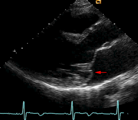
Resumen
Definición
Anamnesis y examen
Principales factores de diagnóstico
- midsystolic click
- late-systolic murmur
Otros factores de diagnóstico
- palpitations
Factores de riesgo
- family history
- slim body type
- connective tissue disorders
- mitral annular disjunction (MAD)
Pruebas diagnósticas
Primeras pruebas diagnósticas para solicitar
- echocardiogram
Pruebas diagnósticas que deben considerarse
- Holter or event monitor
- cardiac MRI
Algoritmo de tratamiento
asymptomatic
symptomatic
Colaboradores
Autores
Sheila Klassen, MD
Assistant Professor of Medicine
Harvard Medical School
Associate Director of Cardiovascular Integration
Center for Integration Science
Brigham and Women's Hospital
Boston
MA
Divulgaciones
SK declares that he has no competing interests.
Agradecimientos
Dr Sheila Klassen would like to gratefully acknowledge Dr Brian Griffin, Dr Shaberio Lo Presti, Dr Carmel Halley, and Dr Serge Harb, previous contributors to this topic.
Disclosures
BG, SLP, CH, and SH declared that they had no competing interests.
Revisores por pares
John Dent, MD
Associate Professor of Internal Medicine
Director of Stress Testing Laboratory and Echocardiography
University of Virginia Health System
Cardiovascular Fellowship Program
Charlottesville
VA
Divulgaciones
JD declares that he has no competing interests.
Prakash Punjabi, MBBS
Honorary Clinical Senior Lecturer
National Heart and Lung Institute
Imperial College London
London
UK
Divulgaciones
PP declares that he has no competing interests.
Agradecimiento de los revisores por pares
Los temas de BMJ Best Practice se actualizan de forma continua de acuerdo con los desarrollos en la evidencia y en las guías. Los revisores por pares listados aquí han revisado el contenido al menos una vez durante la historia del tema.
Divulgaciones
Las afiliaciones y divulgaciones de los revisores por pares se refieren al momento de la revisión.
Referencias
Artículos principales
Otto CM, Nishimura RA, Bonow RO, et al. 2020 ACC/AHA guideline for the management of patients with valvular heart disease: a report of the American College of Cardiology/American Heart Association Joint Committee on Clinical Practice Guidelines. Circulation. 2021 Feb 2;143(5):e72-e227.Texto completo Resumen
Delgado V, Ajmone Marsan N, de Waha S, et al. 2023 ESC guidelines for the management of endocarditis. Eur Heart J. 2023 Oct 14;44(39):3948-4042.Texto completo
Vahanian A, Beyersdorf F, Praz F, et al. 2021 ESC/EACTS guidelines for the management of valvular heart disease. Eur Heart J. 2021 Aug 28:ehab395.Texto completo Resumen
Artículos de referencia
Una lista completa de las fuentes a las que se hace referencia en este tema está disponible para los usuarios con acceso a todo BMJ Best Practice.

Diferenciales
- Aortic valve disease
- Pulmonic valve disease
- Atrial myxoma
Más DiferencialesGuías de práctica clínica
- 2023 ESC guidelines for the management of endocarditis
- 2021 ESC/EACTS guidelines for the management of valvular heart disease
Más Guías de práctica clínicaFolletos para el paciente
Mitral valve prolapse
Más Folletos para el pacienteInicie sesión o suscríbase para acceder a todo el BMJ Best Practice
El uso de este contenido está sujeto a nuestra cláusula de exención de responsabilidad