Summary
Definition
IE is an infection involving the endocardial surface of the heart, including the valvular structures, the chordae tendineae, sites of septal defects, or the mural endocardium.[1][Figure caption and citation for the preceding image starts]: Gross pathology of subacute bacterial endocarditis involving mitral valveCDC/Dr Edwin P. Ewing, Jr.; used with permission [Citation ends].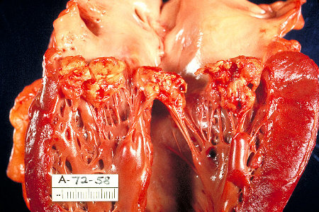 Chronic IE is not covered in this topic.
History and exam
Key diagnostic factors
- fever/chills
- night sweats, malaise, fatigue, anorexia, weight loss, myalgias
- weakness
- arthralgias
- headache
- shortness of breath
- meningeal signs
- cardiac murmur
- Janeway lesions
- Osler nodes
- Roth spots
Other diagnostic factors
- splinter hemorrhages
- cutaneous infarcts
- chest pain
- back pain
- palatal petechiae
Risk factors
- prior history of infectious endocarditis
- presence of artificial prosthetic heart valves
- certain types of congenital heart disease
- postheart transplant (patients who develop a cardiac valvulopathy)
- immunocompromised state
- presence of cardiac implanted electronic device or intravascular catheters (e.g., for hemodialysis)
- acquired degenerative valve disease
- mitral valve prolapse or bicuspid valve prolapse
- hypertrophic cardiomyopathy
- intravenous drug use
- chronic Q fever
Diagnostic tests
1st tests to order
- blood cultures
- echocardiogram
- CBC
- CRP
- serum chemistry panel with glucose
- LFTs
- urinalysis
- ECG
Tests to consider
- rheumatoid factor
- erythrocyte sedimentation rate
- complement levels
- cardiac CT
- MRI
- nuclear imaging and PET
- specific tests for blood culture-negative endocarditis
Emerging tests
- mean platelet volume (MPV)
- anti‐beta-2‐glycoprotein I antibodies
- D-dimer and troponin I
Treatment algorithm
suspected infective endocarditis
native valve: confirmed endocarditis
prosthetic valve: confirmed endocarditis
at high risk of infective endocarditis
Contributors
Expert advisers
Carl Zehner, MD
Assistant Professor of Medicine
University of Texas MD Anderson Cancer Center
Department of Cardiology
Houston, TX
Disclosures
CZ declares that he has no competing interests.
Acknowledgements
Dr Carl Zehner would like to gratefully acknowledge Dr Ammara Mushtuq, Dr Tracey Keteepe-Arachi, Dr Aneil Malhotra, Dr Michael Papadakis, Professor Sanjay Sharma, Dr Jason C. Schultz, Dr Nisha K. Lassi, and Dr Nandan S. Anavekar, previous contributors to this topic.
Disclosures
TKA, MP, SS, JCS, NKL, and NSA declare that they have no competing interests. AMu declares that she is Principal Investigator on a grant from Merck on ceftolozane-tazobactam, writer for the Lancet family of journals, question writer for Blueprint Test preparations, and a peer-reviewer for Clinical Overviews, Elsevier (topics influenza and HIV). AMa is an author of a number of references cited in this topic.
Peer reviewers
Vandana Desai, MD
Professor of Pediatrics
SMIMER Hospital
Gujarat
India
Disclosures
VD declares that she has no competing interests.
Lucieni Oliveira Conterno, MD, PhD
Director
Clinical Epidemiology Unit
Marilia Medical School
Sao Paulo
Brazil
Disclosures
LOC declares that she has no competing interests.
Andrew Wang, MD
Associate Professor of Medicine
Director, Cardiovascular Disease Fellowship Program
Duke University Medical Center
Durham, NC
Disclosures
AW declares that he has no competing interests.
Peer reviewer acknowledgements
BMJ Best Practice topics are updated on a rolling basis in line with developments in evidence and guidance. The peer reviewers listed here have reviewed the content at least once during the history of the topic.
Disclosures
Peer reviewer affiliations and disclosures pertain to the time of the review.
References
Key articles
Baddour LM, Wilson WR, Bayer AS, et al. Infective endocarditis in adults: diagnosis, antimicrobial therapy, and management of complications: a scientific statement for healthcare professionals from the American Heart Association. Circulation. 2015 Oct 13;132(15):1435-86.Full text Abstract
Delgado V, Ajmone Marsan N, de Waha S, et al. 2023 ESC guidelines for the management of endocarditis. Eur Heart J. 2023 Oct 14;44(39):3948-4042.Full text Abstract
Otto CM, Nishimura RA, Bonow RO, et al. 2020 ACC/AHA guideline for the management of patients with valvular heart disease: a report of the American College of Cardiology/American Heart Association Joint Committee on clinical practice guidelines. Circulation. 2021 Feb 2;143(5):e72-227.Full text Abstract
Wilson WR, Gewitz M, Lockhart PB, et al. Prevention of viridans group streptococcal infective endocarditis: a scientific statement from the American Heart Association. Circulation. 2021 May 18;143(20):e963-78.Full text Abstract
Reference articles
A full list of sources referenced in this topic is available to users with access to all of BMJ Best Practice.
Differentials
- Rheumatic fever
- Atrial myxoma
- Libman-Sacks endocarditis
More DifferentialsGuidelines
- Blood culture-negative endocarditis
- 2023 ESC guidelines for the management of endocarditis
More GuidelinesVideos
Venepuncture and phlebotomy: animated demonstration
How to perform an ECG: animated demonstration
More videosPatient information
Endocarditis
More Patient information Log in or subscribe to access all of BMJ Best Practice
Use of this content is subject to our disclaimer