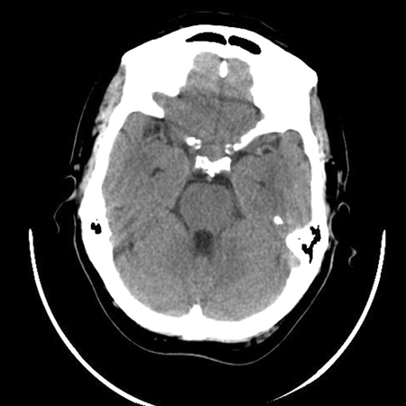
Summary
Differentials
Common
- Prolactinoma
- Nonfunctional adenoma
- Gonadotropin-secreting adenoma
- Lactotroph hyperplasia
Uncommon
- Adrenocorticotropic hormone (ACTH)-secreting adenoma
- Growth hormone (GH)-secreting adenoma
- Thyroid-stimulating hormone (TSH)-secreting adenoma
- Thyrotroph hyperplasia
- Gonadotroph hyperplasia
- Somatotroph hyperplasia
- Corticotroph hyperplasia
- Craniopharyngioma
- Meningioma
- Germ cell tumors
- Lymphoma
- Chordoma
- Metastatic disease
- Lymphocytic hypophysitis
- Drug therapy-induced hypophysitis
- Pituitary abscess
- Pituitary apoplexy
- Cerebral aneurysm
- Rathke cleft cyst
- Empty sella syndrome
- Pituitary carcinoma
Contributors
Authors
Joe M. Chehade, MD
Professor of Medicine
Division of Endocrinology
University of Florida
Jacksonville
FL
Disclosures
JMC declares that he has no competing interests.
Acknowledgements
Dr Joe M. Chehade would like to gratefully acknowledge Dr Senan Sultan, a previous contributor to this topic. SS declares that he has no competing interests.
Peer reviewers
Federico Roncaroli, MD
Reader in Neuropathology and Honorary Consultant in Neuropathology
Neuropathology Unit
Department of Clinical Neuroscience
Division of Neuroscience and Mental Health
Faculty of Medicine
Imperial College
London
UK
Disclosures
FR declares that he has no competing interests.
Tony Heaney, MD, PhD
Co-Director
Pituitary Tumor and Neuroendocrine Program
Associate Professor
Medicine and Neurosurgery
David Geffen School of Medicine at UCLA
Los Angeles
CA
Disclosures
TH declares that he has no competing interests.
Peer reviewer acknowledgements
BMJ Best Practice topics are updated on a rolling basis in line with developments in evidence and guidance. The peer reviewers listed here have reviewed the content at least once during the history of the topic.
Disclosures
Peer reviewer affiliations and disclosures pertain to the time of the review.
References
Key articles
Tritos NA, Miller KK. Diagnosis and management of pituitary adenomas: a review. JAMA. 2023 Apr 25;329(16):1386-98. Abstract
Freda PU, Beckers AM, Katznelson L, et al. Pituitary incidentaloma: an Endocrine Society clinical practice guideline. J Clin Endocrinol Metab. 2011;96:894-904.Full text Abstract
American College of Radiology. ACR appropriateness criteria. Neuroendocrine Imaging. 2018 [internet publication].Full text
Reference articles
A full list of sources referenced in this topic is available to users with access to all of BMJ Best Practice.
Use of this content is subject to our disclaimer

