Monoclonal gammopathies represent a wide spectrum of related diseases.[1]Rajkumar SV, Dispenzieri A, Kyle RA. Monoclonal gammopathy of undetermined significance, Waldenstrom macroglobulinemia, AL amyloidosis, and related plasma cell disorders: diagnosis and treatment. Mayo Clin Proc. 2006 May;81(5):693-703.
http://www.ncbi.nlm.nih.gov/pubmed/16706268?tool=bestpractice.com
[2]Kyle RA. Current concepts on monoclonal gammopathies. Aust N Z J Med. 1992 Jun;22(3):291-302.
http://www.ncbi.nlm.nih.gov/pubmed/1497556?tool=bestpractice.com
[3]Kyle RA, Therneau TM, Rajkumar SV, et al. Prevalence of monoclonal gammopathy of undetermined significance. N Engl J Med. 2006 Mar 30;354(13):1362-9.
https://www.nejm.org/doi/full/10.1056/NEJMoa054494
http://www.ncbi.nlm.nih.gov/pubmed/16571879?tool=bestpractice.com
[4]International Myeloma Working Group. Criteria for the classification of monoclonal gammopathies, multiple myeloma and related disorders: a report of the International Myeloma Working Group. Br J Haematol. 2003 Jun;121(5):749-57.
http://www.ncbi.nlm.nih.gov/pubmed/12780789?tool=bestpractice.com
The common denominator is the presence of a monoclonal protein in the serum or urine, which can be in the form of intact immunoglobulin, immunoglobulin fragments, and/or free light chains. This will be accompanied by the presence of monoclonal plasma cells in the bone marrow, in soft tissue (with plasmacytomas), or in the peripheral circulation (in more advanced disease stages).
Plasma cells and monoclonal proteins
Plasma cells are terminally differentiated (specialized cells that typically do not proliferate) effector cells of B-cell lineage.[5]Fairfax KA, Kallies A, Nutt SL, et al. Plasma cell development: from B-cell subsets to long-term survival niches. Semin Immunol. 2008 Feb;20(1):49-58.
http://www.ncbi.nlm.nih.gov/pubmed/18222702?tool=bestpractice.com
[6]McHeyzer-Williams LJ, McHeyzer-Williams MG. Antigen-specific memory B cell development. Annu Rev Immunol. 2005;23:487-513.
http://www.ncbi.nlm.nih.gov/pubmed/15771579?tool=bestpractice.com
[7]Radbruch A, Muehlinghaus G, Luger EO, et al. Competence and competition: the challenge of becoming a long-lived plasma cell. Nat Rev Immunol. 2006 Oct;6(10):741-50.
http://www.ncbi.nlm.nih.gov/pubmed/16977339?tool=bestpractice.com
[8]Shapiro-Shelef M, Calame K. Regulation of plasma-cell development. Nat Rev Immunol. 2005 Mar;5(3):230-42.
http://www.ncbi.nlm.nih.gov/pubmed/15738953?tool=bestpractice.com
They are the primary mediators of humoral immunity, secreting antigen-specific immunoglobulins. Abnormalities of plasma cells are responsible for a variety of autoimmune diseases and plasma cell neoplasms. Clonal evolution of one or more plasma cells sets the stage for development of the monoclonal gammopathies.[5]Fairfax KA, Kallies A, Nutt SL, et al. Plasma cell development: from B-cell subsets to long-term survival niches. Semin Immunol. 2008 Feb;20(1):49-58.
http://www.ncbi.nlm.nih.gov/pubmed/18222702?tool=bestpractice.com
[6]McHeyzer-Williams LJ, McHeyzer-Williams MG. Antigen-specific memory B cell development. Annu Rev Immunol. 2005;23:487-513.
http://www.ncbi.nlm.nih.gov/pubmed/15771579?tool=bestpractice.com
[7]Radbruch A, Muehlinghaus G, Luger EO, et al. Competence and competition: the challenge of becoming a long-lived plasma cell. Nat Rev Immunol. 2006 Oct;6(10):741-50.
http://www.ncbi.nlm.nih.gov/pubmed/16977339?tool=bestpractice.com
[8]Shapiro-Shelef M, Calame K. Regulation of plasma-cell development. Nat Rev Immunol. 2005 Mar;5(3):230-42.
http://www.ncbi.nlm.nih.gov/pubmed/15738953?tool=bestpractice.com
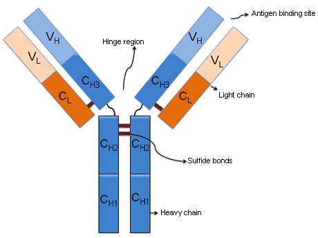
Plasma cells normally secrete an intact immunoglobulin that is made up of 2 identical light chains and 2 identical heavy chains. There are 5 major classes of heavy chains, which correspond to the major classes of immunoglobulins: mu (IgM), delta (IgD), gamma (IgG), alpha (IgA), and IgE (epsilon). In each of the immunoglobulin molecules, the heavy chains are bound to one or the other of the 2 light chains (either kappa or lambda), but not both. The heavy chains, which have 4 or 5 domains, and the light chains, which have 2 domains, are covalently bonded to each other through disulfide bonds.[Figure caption and citation for the preceding image starts]: Immunoglobulin structureFrom the personal collection of Dr Kumar [Citation ends].
Monoclonal proteins are abnormal, immunologically homogeneous immunoglobulins or parts of immunoglobulins, produced by a single clone of plasma cells. They may be the result of an underlying lymphoid malignancy, be part of a clonal expansion of plasma cells causing no symptoms (e.g., monoclonal gammopathy of unknown significance), or lead to life-threatening complications (e.g., primary amyloidosis).[9]Kumar S, Dispenzieri A, Katzmann JA, et al. Serum immunoglobulin free light-chain measurement in primary amyloidosis: prognostic value and correlations with clinical features. Blood. 2010 Dec 9;116(24):5126-9.
http://bloodjournal.hematologylibrary.org/content/116/24/5126.long
http://www.ncbi.nlm.nih.gov/pubmed/20798235?tool=bestpractice.com
Protein electrophoresis is performed to detect and identify monoclonal proteins in the serum and urine.[10]Katzmann JA, Kyle R. Immunochemical characterization of immunoglobulins in serum, urine, and cerebrospinal fluid. In: Detrick B, ed. Manual of molecular and clinical laboratory immunology. 7th ed. Washington, DC: American Society for Microbiology Press; 2006:88-100.[11]Katzmann JA, Dispenzieri A. Screening algorithms for monoclonal gammopathies. Clin Chem. 2008 Nov;54(11):1753-5.
http://www.clinchem.org/cgi/content/full/54/11/1753
http://www.ncbi.nlm.nih.gov/pubmed/18957556?tool=bestpractice.com
Quantitation of immunoglobulins is achieved by nephelometry. Immunoglobulin free light chains in serum are assessed using antibodies specific for the light chain part of the immunoglobulins.
Clonal expansion of plasma cells
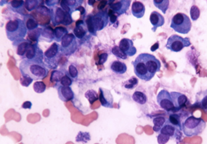
Clonal expansion of plasma cells is the underlying abnormality among the monoclonal gammopathies. These cells may be found in the bone marrow, peripheral circulation, or soft tissue. They are typically demonstrated on bone marrow exams, where presence of clonal plasma cells may or may not be accompanied by an absolute increase in the plasma cell proportion. Whereas plasma cells are identified by their surface staining for CD138 (syndecan) on immunohistochemistry, demonstration of clonality depends on light chain restriction, and an excess of kappa- or lambda-expressing plasma cells resulting in a skewing of the normal kappa to lambda ratio.[12]O'Connell FP, Pinkus JL, Pinkus GS. CD138 (syndecan-1), a plasma cell marker immunohistochemical profile in hematopoietic and nonhematopoietic neoplasms. Am J Clin Pathol. 2004 Feb;121(2):254-63.
http://www.ncbi.nlm.nih.gov/pubmed/14983940?tool=bestpractice.com
The skewed ratio can be demonstrated by immunohistochemistry using antibodies against kappa or lambda light chains or by polymerase chain reaction (PCR) performed on marrow biopsy specimens.
Clonal plasma cells numbers are typically small in most monoclonal gammopathies, except for advanced myeloma and plasma cell leukemia.[13]Witzig TE, Kimlinger TK, Ahmann GJ, et al. Detection of myeloma cells in the peripheral blood by flow cytometry. Cytometry. 1996 Jun 15;26(2):113-20.
http://www.ncbi.nlm.nih.gov/pubmed/8817086?tool=bestpractice.com
[14]Albarracin F, Fonseca R. Plasma cell leukemia. Blood Rev. 2011 May;25(3):107-12.
http://www.ncbi.nlm.nih.gov/pubmed/21295388?tool=bestpractice.com
Sensitive flow cytometry techniques can be used for routine evaluation of bone marrow aspirates to detect small numbers and for immunophenotypic characterization of abnormal plasma cells.[15]Lin P, Owens R, Tricot G, et al. Flow cytometric immunophenotypic analysis of 306 cases of multiple myeloma. Am J Clin Pathol. 2004 Apr;121(4):482-8.
http://www.ncbi.nlm.nih.gov/pubmed/15080299?tool=bestpractice.com
[16]Paiva B, Vidriales MB, Cervero J, et al. Multiparameter flow cytometric remission is the most relevant prognostic factor for multiple myeloma patients who undergo autologous stem cell transplantation. Blood. 2008 Nov 15;112(10):4017-23.
https://www.ncbi.nlm.nih.gov/pmc/articles/PMC2581991
http://www.ncbi.nlm.nih.gov/pubmed/18669875?tool=bestpractice.com
[17]Rawstron AC, Orfao A, Beksac M, et al. Report of the European Myeloma Network on multiparametric flow cytometry in multiple myeloma and related disorders. Haematologica. 2008 Mar;93(3):431-8.
https://haematologica.org/article/download/4784/18859
http://www.ncbi.nlm.nih.gov/pubmed/18268286?tool=bestpractice.com
[Figure caption and citation for the preceding image starts]: Monoclonal plasma cellsFrom the personal collection of Dr Kumar [Citation ends].
Epidemiology
Monoclonal gammopathy of undetermined significance (MGUS) is the most common monoclonal gammopathy and is found in about 1% to 2% of adults.[3]Kyle RA, Therneau TM, Rajkumar SV, et al. Prevalence of monoclonal gammopathy of undetermined significance. N Engl J Med. 2006 Mar 30;354(13):1362-9.
https://www.nejm.org/doi/full/10.1056/NEJMoa054494
http://www.ncbi.nlm.nih.gov/pubmed/16571879?tool=bestpractice.com
[18]Axelsson U, Hallen J. Review of fifty-four subjects with monoclonal gammopathy. Br J Haematol. 1968 Oct;15(4):417-20.
http://www.ncbi.nlm.nih.gov/pubmed/4176071?tool=bestpractice.com
[19]Kyle RA, Therneau TM, Rajkumar SV, et al. A long-term study of prognosis in monoclonal gammopathy of undetermined significance. N Engl J Med. 2002 Feb 21;346(8):564-9.
https://www.nejm.org/doi/full/10.1056/NEJMoa01133202#t=article
http://www.ncbi.nlm.nih.gov/pubmed/11856795?tool=bestpractice.com
[20]Saleun JP, Vicariot M, Deroff P, et al. Monoclonal gammopathies in the adult population of Finistere, France. J Clin Pathol. 1982 Jan;35(1):63-8.
https://www.ncbi.nlm.nih.gov/pmc/articles/PMC497449
http://www.ncbi.nlm.nih.gov/pubmed/6801095?tool=bestpractice.com
[21]Kyle RA, Durie BG, Rajkumar SV, et al; International Myeloma Working Group. Monoclonal gammopathy of undetermined significance (MGUS) and smoldering (asymptomatic) multiple myeloma: IMWG consensus perspectives risk factors for progression and guidelines for monitoring and management. Leukemia. 2010 Jun;24(6):1121-7.
http://www.ncbi.nlm.nih.gov/pubmed/20410922?tool=bestpractice.com
[22]Wadhera RK, Rajkumar SV. Prevalence of monoclonal gammopathy of undetermined significance: a systematic review. Mayo Clin Proc. 2010 Oct;85(10):933-42.
http://www.ncbi.nlm.nih.gov/pubmed/20713974?tool=bestpractice.com
The prevalence increases with age and is generally higher in males compared with females, and in patients over 70 years of age.[3]Kyle RA, Therneau TM, Rajkumar SV, et al. Prevalence of monoclonal gammopathy of undetermined significance. N Engl J Med. 2006 Mar 30;354(13):1362-9.
https://www.nejm.org/doi/full/10.1056/NEJMoa054494
http://www.ncbi.nlm.nih.gov/pubmed/16571879?tool=bestpractice.com
[23]Kyle RA, Rajkumar SV. Monoclonal gammopathies of undetermined significance. Best Pract Res Clin Haematol. 2005;18(4):689-707.
https://www.doi.org/10.1016/j.beha.2005.01.025
http://www.ncbi.nlm.nih.gov/pubmed/16026745?tool=bestpractice.com
Prevalence rates vary according to geographic and racial factors, with lower rates in Asia compared with those in Europe and North and South America, and higher prevalence rates among black people compared with white people.[24]Landgren O, Katzmann JA, Hsing AW, et al. Prevalence of monoclonal gammopathy of undetermined significance among men in Ghana. Mayo Clin Proc. 2007 Dec;82(12):1468-73.
http://www.ncbi.nlm.nih.gov/pubmed/18053453?tool=bestpractice.com
Increased prevalence has been observed among first-degree relatives of patients with monoclonal gammopathies.[25]Vachon CM, Kyle RA, Therneau TM, et al. Increased risk of monoclonal gammopathy in first-degree relatives of patients with multiple myeloma or monoclonal gammopathy of undetermined significance. Blood. 2009 Jul 23;114(4):785-90.
https://www.ncbi.nlm.nih.gov/pmc/articles/PMC2716020
http://www.ncbi.nlm.nih.gov/pubmed/19179466?tool=bestpractice.com
Associated conditions
There are disease conditions other than MGUS where a monoclonal protein may be detected in the serum and/or urine. These include:
Lymphoproliferative diseases where clonal B lineage cells secrete a monoclonal protein (chronic lymphocytic leukemia, non-Hodgkin lymphoma, post-transplant monoclonal gammopathies)[26]Wadhera RK, Kyle RA, Larson DR, et al. Incidence, clinical course, and prognosis of secondary monoclonal gammopathy of undetermined significance in patients with multiple myeloma. Blood. 2011 Sep 15;118(11):2985-7.
http://bloodjournal.hematologylibrary.org/content/early/2011/07/15/blood-2011-04-349175.long
http://www.ncbi.nlm.nih.gov/pubmed/21765020?tool=bestpractice.com
Conditions associated with or predisposing to a higher prevalence of monoclonal gammopathy (hepatitis C virus infection, HIV infection)
Infectious or inflammatory conditions associated with a transient development of several clones of reactive B cell/plasma cell populations (systemic lupus erythematosus, rheumatoid arthritis, psoriatic arthritis, Sjogren syndrome, Schnitzler syndrome).

Log in or subscribe to access all of BMJ Best Practice
