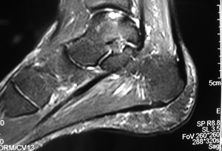
Summary
Definition
History and exam
Key diagnostic factors
- presence of risk factors
- pain is exacerbated by activity
- location of pain anteromedial aspect of the knee with the knee flexed to 90º
- location of pain lateral aspect of elbow
- location of pain posteromedial aspect of dorsiflexed ankle or anterolateral aspect of plantar-flexed ankle
- effusion present
- locking of joint
- catching of joint
- decreased range of motion
Other diagnostic factors
- knee involvement, age 10 to 20 years
- elbow involvement, age 11 to 21 years
- talus involvement, second to fourth decade
- absence of history of trauma involving the knee or elbow
- antalgic gait in osteochondritis dissecans involving the knee or talus
- external rotation gait in osteochondritis dissecans involving the knee
- relieving factors: non-steroidal anti-inflammatory drugs (NSAIDS), rest, ice, elevation
- crepitus
- Wilson's test
- quadriceps atrophy
Risk factors
- repetitive throwing/valgus stress
- gymnastics/weight-bearing on upper extremity
- ankle sprain/instability
- competitive athletics
- family history
Diagnostic investigations
1st investigations to order
- knee x-rays
- ankle x-rays
- full-length lower extremity film
- elbow x-rays
Investigations to consider
- CT
- MRI
- MR arthrogram
- diagnostic arthroscopy
Treatment algorithm
knee
elbow
ankle (talus)
Contributors
Authors
Henry G. Chambers, MD
Professor of Clinical Orthopedic Surgery
University of California, San Diego
Rady Children’s Hospital
San Diego
CA
Disclosures
HGC is an author of a number of references cited in this topic.
Acknowledgements
Dr Henry G. Chambers would like to gratefully acknowledge Dr James L. Carey, Dr Jon Divine, Dr Michael Nett, and Dr Cedric Ortiguera, the previous contributors to this topic.
Disclosures
JLC is an author of a number of references cited in this topic. JD, MN, and CO declared that they had no competing interests.
Peer reviewers
James E. McGrory, MD
Orthopedic Surgeon
The Hughston Clinic PC
Columbus
GA
Disclosures
JEM declares that he has no competing interests.
Nicola Maffulli, MD, MS, PhD, FRCS(Orth)
Centre Lead and Professor of Sports and Exercise Medicine
Consultant Trauma and Orthopaedic Surgeon
Barts and The London School of Medicine and Dentistry
Institute for Health Sciences Education
Centre for Sports and Exercise Medicine
Queen Mary University of London
Mile End Hospital
London
UK
Disclosures
NM declares that he has no competing interests.
Peer reviewer acknowledgements
BMJ Best Practice topics are updated on a rolling basis in line with developments in evidence and guidance. The peer reviewers listed here have reviewed the content at least once during the history of the topic.
Disclosures
Peer reviewer affiliations and disclosures pertain to the time of the review.
References
Key articles
Kocher MS, Tucker R, Ganley TJ, et al. Management of osteochondritis dissecans of the knee: current concepts review. Am J Sports Med. 2006 Jul;34(7):1181-91. Abstract
American Academy of Orthopaedic Surgeons. Diagnosis and treatment of osteochondritis dissecans. Dec 2023 [internet publication].Full text
Perumal V, Wall E, Babekir N. Juvenile osteochondritis dissecans of the talus. J Pediatr Orthop. 2007 Oct-Nov;27(7):821-5. Abstract
Baker CL 3rd, Baker CL Jr, Romeo AA. Osteochondritis dissecans of the capitellum. Am J Sports Med. 2010 Sep;38(9):1917-28. Abstract
Reference articles
A full list of sources referenced in this topic is available to users with access to all of BMJ Best Practice.

Differentials
- Osteochondral fracture
- Meniscal tear
- Septic arthritis
More DifferentialsGuidelines
- Osteochondritis dissecans: diagnosis and treatment
More GuidelinesLog in or subscribe to access all of BMJ Best Practice
Use of this content is subject to our disclaimer