The signs and symptoms of bacterial meningitis depend on the patient's age. In children particularly, early signs and symptoms of meningitis can be nonspecific and similar to other common, less serious illnesses. Infants may present with very nonspecific signs, such as tachypnea, bulging fontanelle, irritability, or back arching.[53]Chávez-Bueno S, McCracken GH Jr. Bacterial meningitis in children. Pediatr Clin North Am. 2005 Jun;52(3):795-810.
http://www.ncbi.nlm.nih.gov/pubmed/15925663?tool=bestpractice.com
[54]Boston Children's Hospital. Bacterial meningitis [internet publication].
https://www.childrenshospital.org/conditions-treatments/bacterial-meningitis-children
[55]Skar G, Flannigan L, Latch R, et al. Meningitis in children: still a can't-miss diagnosis. Pediatr Rev. 2024 Jun 1;45(6):305-15.
http://www.ncbi.nlm.nih.gov/pubmed/38821894?tool=bestpractice.com
Even when a diagnosis of bacterial meningitis appears unlikely at the time of presentation, information provided to patients, parents, or caregivers should include:[56]National Institute for Health and Care Excellence. Fever in under 5s: assessment and initial management. Nov 2021 [internet publication].
https://www.nice.org.uk/guidance/ng143
[57]National Institute for Health and Care Excellence. Meningitis (bacterial) and meningococcal disease: recognition, diagnosis and management. Mar 2024 [internet publication].
https://www.nice.org.uk/guidance/ng240/chapter/Recommendations
[58]World Health Organization. WHO guidelines on meningitis diagnosis, treatment and care. Apr 2025 [internet publication].
https://www.who.int/publications/i/item/9789240108042
It may be impossible to differentiate between viral and bacterial meningitis clinically. The diagnosis is confirmed by exam and polymerase chain reaction (PCR), culture of cerebrospinal fluid (CSF) obtained from a lumbar puncture (LP), or blood culture (if an LP is not clinically safe to obtain).
History
Classic symptoms of meningitis in children and adults include fever, severe headache, neck stiffness, photophobia, altered mental status, vomiting, and seizures.[1]Mace SE. Acute bacterial meningitis. Emerg Med Clin North Am. 2008 May;26(2):281-317.
http://www.ncbi.nlm.nih.gov/pubmed/18406976?tool=bestpractice.com
[9]Sáez-Llorens X, McCracken GH Jr. Bacterial meningitis in children. Lancet. 2003 Jun 21;361(9375):2139-48.
http://www.ncbi.nlm.nih.gov/pubmed/12826449?tool=bestpractice.com
The classic triad of fever, neck stiffness, and altered mental status occurs in only 41% to 51% of patients.[59]van de Beek D, Cabellos C, Dzupova O, et al. ESCMID guideline: diagnosis and treatment of acute bacterial meningitis. Clin Microbiol Infect. 2016 May;22(suppl 3):S37-62.
https://www.clinicalmicrobiologyandinfection.com/article/S1198-743X(16)00020-3/fulltext
http://www.ncbi.nlm.nih.gov/pubmed/27062097?tool=bestpractice.com
However, in one study, 95% had at least two of the four symptoms of headache, fever, neck stiffness, and altered mental status.[42]van de Beek D, de Gans J, Spanjaard L, et al. Clinical features and prognostic factors in adults with bacterial meningitis. N Engl J Med. 2004 Oct 28;351(18):1849-59.
https://www.nejm.org/doi/full/10.1056/NEJMoa040845
http://www.ncbi.nlm.nih.gov/pubmed/15509818?tool=bestpractice.com
Children have seizures more frequently with meningitis caused by Streptococcus pneumoniae and Haemophilus influenzae type b (Hib) than with meningococcal meningitis.[1]Mace SE. Acute bacterial meningitis. Emerg Med Clin North Am. 2008 May;26(2):281-317.
http://www.ncbi.nlm.nih.gov/pubmed/18406976?tool=bestpractice.com
Atypical clinical manifestations tend to occur in very young, older, or immunocompromised patients. In infants, the signs and symptoms can be nonspecific and may include fever, hypothermia, irritability, high-pitched crying, lethargy, poor feeding, seizures, apnea, or a bulging fontanel.[53]Chávez-Bueno S, McCracken GH Jr. Bacterial meningitis in children. Pediatr Clin North Am. 2005 Jun;52(3):795-810.
http://www.ncbi.nlm.nih.gov/pubmed/15925663?tool=bestpractice.com
Frequently in older patients (>65 years), the only presenting sign of meningitis is confusion or an altered mental status.[1]Mace SE. Acute bacterial meningitis. Emerg Med Clin North Am. 2008 May;26(2):281-317.
http://www.ncbi.nlm.nih.gov/pubmed/18406976?tool=bestpractice.com
Focal neurologic deficits, including aphasia, hemiparesis, or cranial nerve palsies, may be present in adults with community-acquired bacterial meningitis.[60]Bijlsma MW, Brouwer MC, Kasanmoentalib ES, et al. Community-acquired bacterial meningitis in adults in the Netherlands, 2006-14: a prospective cohort study. Lancet Infect Dis. 2016 Mar;16(3):339-47.
http://www.ncbi.nlm.nih.gov/pubmed/26652862?tool=bestpractice.com
If the disease has been allowed to progress, the patient may present with signs of sepsis, such as decreased peripheral perfusion, tachypnea, or rapidly evolving petechial or purpuric rash.[7]World Health Organization. Meningitis. Apr 2025 [internet publication].
https://www.who.int/news-room/fact-sheets/detail/meningitis#:~:text=These%20bacteria%20are%20responsible%20for,also%20important%20causes%20of%20meningitis
[61]Meningitis Research Foundation. What is the 'meningitis rash'? Mar 2019 [internet publication].
https://www.meningitis.org/blogs/what-is-the-meningitis-rash
A late diagnosis of bacterial meningitis is associated with higher mortality.[62]Bodilsen J, Brandt CT, Sharew A, et al. Early versus late diagnosis in community-acquired bacterial meningitis: a retrospective cohort study. Clin Microbiol Infect. 2018 Feb;24(2):166-70.
https://www.clinicalmicrobiologyandinfection.com/article/S1198-743X(17)30345-2/fulltext
http://www.ncbi.nlm.nih.gov/pubmed/28652113?tool=bestpractice.com
A careful history to rule out possible viral infections, such as enteroviruses (e.g., other sick children or family members) or herpes virus infection (e.g., sore on lip or genital lesions), should be taken. Immunization history against H influenzae type b, S pneumoniae, and Neisseria meningitidis should be established.
Exam
After assessment of vital signs and mental status, the following should be evaluated:
Nuchal rigidity
A stiff neck with resistance to passive neck flexion is a classic sign of meningitis. It is present in 83% of adults with bacterial meningitis, but may be present in only 30% of children.[42]van de Beek D, de Gans J, Spanjaard L, et al. Clinical features and prognostic factors in adults with bacterial meningitis. N Engl J Med. 2004 Oct 28;351(18):1849-59.
https://www.nejm.org/doi/full/10.1056/NEJMoa040845
http://www.ncbi.nlm.nih.gov/pubmed/15509818?tool=bestpractice.com
[59]van de Beek D, Cabellos C, Dzupova O, et al. ESCMID guideline: diagnosis and treatment of acute bacterial meningitis. Clin Microbiol Infect. 2016 May;22(suppl 3):S37-62.
https://www.clinicalmicrobiologyandinfection.com/article/S1198-743X(16)00020-3/fulltext
http://www.ncbi.nlm.nih.gov/pubmed/27062097?tool=bestpractice.com
[63]Oostenbrink R, Moons KG, Theunissen CC, et al. Signs of meningeal irritation at the emergency department: how often bacterial meningitis? Pediatr Emerg Care. 2001 Jun;17(3):161-4.
http://www.ncbi.nlm.nih.gov/pubmed/11437138?tool=bestpractice.com
Rash
A petechial or purpuric rash is typically associated with meningococcal meningitis but can also occur in pneumococcal meningitis.[42]van de Beek D, de Gans J, Spanjaard L, et al. Clinical features and prognostic factors in adults with bacterial meningitis. N Engl J Med. 2004 Oct 28;351(18):1849-59.
https://www.nejm.org/doi/full/10.1056/NEJMoa040845
http://www.ncbi.nlm.nih.gov/pubmed/15509818?tool=bestpractice.com
Although only a few patients with fever and a petechial rash will ultimately be found to have meningococcal infections, these findings should prompt investigations to exclude meningococcemia and to start empiric antibacterial therapy unless an alternative diagnosis is likely.
Papilledema, bulging fontanel in infants
Evidence of primary source of infection
The patient may also have sinusitis, pneumonia, mastoiditis, or otitis media that suggest pneumococcal meningitis.
Cranial nerve palsy (III, IV, VI)
This is suggested by problems with eye movement and is probably related to increased intracranial pressure.
Cranial nerves VII and VIII may also be involved and damaged due to increased intracranial pressure and inflammation. This damage may lead to facial palsy, balance problems, and hearing impairment.
Kernig and Brudzinski signs
Positive signs are indicators of meningitis commonly in older children and adults, but they may be absent in up to 50% of adults.[1]Mace SE. Acute bacterial meningitis. Emerg Med Clin North Am. 2008 May;26(2):281-317.
http://www.ncbi.nlm.nih.gov/pubmed/18406976?tool=bestpractice.com
Kernig sign: with the patient supine and the thigh flexed to a 90° right angle, attempts to straighten or extend the leg are met with resistance.
Brudzinski signs: flexion of the neck causes involuntary flexion of the knees and hips, or passive flexion of the leg on one side causes contralateral flexion of the opposite leg.
Investigations
LP and analysis of CSF
An LP to obtain CSF is the most important investigation when a diagnosis of bacterial meningitis is suspected; however, if LP is delayed (e.g., due to awaiting imaging), this should not delay initiation of antibiotics.[59]van de Beek D, Cabellos C, Dzupova O, et al. ESCMID guideline: diagnosis and treatment of acute bacterial meningitis. Clin Microbiol Infect. 2016 May;22(suppl 3):S37-62.
https://www.clinicalmicrobiologyandinfection.com/article/S1198-743X(16)00020-3/fulltext
http://www.ncbi.nlm.nih.gov/pubmed/27062097?tool=bestpractice.com
At least 15 mL of CSF is required for investigations.[64]McGill F, Heyderman RS, Michael BD, et al. The UK joint specialist societies guideline on the diagnosis and management of acute meningitis and meningococcal sepsis in immunocompetent adults. J Infect. 2016 Apr;72(4):405-38.
https://www.journalofinfection.com/article/S0163-4453(16)00024-4/fulltext
http://www.ncbi.nlm.nih.gov/pubmed/26845731?tool=bestpractice.com
In bacterial meningitis the CSF pressure is usually elevated (>20 cm H₂O).
The CSF white blood cell count is elevated (usually >1000 cells/microliter), of which over 90% are polymorphonuclear leukocytes. In neonates, however, the CSF leukocyte count may be normal.[65]Garges HP, Moody MA, Cotten CM, et al. Neonatal meningitis: what is the correlation among cerebrospinal fluid cultures, blood cultures, and cerebrospinal fluid parameters? Pediatrics. 2006 Apr;117(4):1094-100.
http://www.ncbi.nlm.nih.gov/pubmed/16585303?tool=bestpractice.com
The glucose level in the CSF is decreased compared with the serum value, and the protein level is increased, indicating accelerated bacterial growth and blood brain barrier disruption. In untreated patients, Gram stain and bacterial culture of the CSF are usually positive for the causative organism. Bacterial culture of CSF is positive in 80% of cases.[42]van de Beek D, de Gans J, Spanjaard L, et al. Clinical features and prognostic factors in adults with bacterial meningitis. N Engl J Med. 2004 Oct 28;351(18):1849-59.
https://www.nejm.org/doi/full/10.1056/NEJMoa040845
http://www.ncbi.nlm.nih.gov/pubmed/15509818?tool=bestpractice.com
However, diagnostic yields can be much lower in patients who have received antibiotics before cultures are obtained. [Figure caption and citation for the preceding image starts]: species bacteriaImage provided by the CDC Public Health Image Library [Citation ends].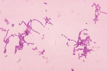
Serogroup A, B, C, Y, and W-135 polysaccharide antigen can be detected by latex agglutination in 22% to 93% of patients with meningococcal meningitis.[4]Brouwer MC, Tunkel AR, van de Beek D. Epidemiology, diagnosis, and antimicrobial treatment of acute bacterial meningitis. Clin Microbiol Rev. 2010 Jul;23(3):467-92.
https://cmr.asm.org/content/23/3/467.long
http://www.ncbi.nlm.nih.gov/pubmed/20610819?tool=bestpractice.com
Antigen may persist in the CSF for several days, making this test useful in patients treated with antibiotics before diagnostic specimens could be obtained, and for the rapid presumptive diagnosis of meningococcal infection. Serogroup B N meningitidis and serotype K1 Escherichia coli polysaccharides cross-react, so test results should be interpreted cautiously in neonates. A rapid pneumococcal antigen in the CSF has shown 99% sensitivity and 99% specificity to rule in or out pneumococcal meningitis in low-income countries.[72]Moïsi JC, Saha SK, Falade AG, et al. Enhanced diagnosis of pneumococcal meningitis with use of the Binax NOW immunochromatographic test of Streptococcus pneumoniae antigen: a multisite study. Clin Infect Dis. 2009 Mar 1;48 Suppl 2(suppl 2):S49-56.
https://pmc.ncbi.nlm.nih.gov/articles/PMC2863072
http://www.ncbi.nlm.nih.gov/pubmed/19191619?tool=bestpractice.com
Antigen detection testing on body fluids other than CSF, including serum or urine, is not recommended because of poor sensitivity and specificity.
A cranial computed tomography (CT) scan should be considered before LP in the presence of focal neurologic deficit, new-onset seizures, papilledema, abnormal level of consciousness, or immunocompromised state to exclude a brain abscess or generalized cerebral edema.[59]van de Beek D, Cabellos C, Dzupova O, et al. ESCMID guideline: diagnosis and treatment of acute bacterial meningitis. Clin Microbiol Infect. 2016 May;22(suppl 3):S37-62.
https://www.clinicalmicrobiologyandinfection.com/article/S1198-743X(16)00020-3/fulltext
http://www.ncbi.nlm.nih.gov/pubmed/27062097?tool=bestpractice.com
PCR
PCR amplification of bacterial DNA from blood and CSF is more sensitive and specific than traditional microbiologic techniques. It is especially useful in distinguishing bacterial from viral meningitis; false-negative results are uncommon (about 5% of cases).[4]Brouwer MC, Tunkel AR, van de Beek D. Epidemiology, diagnosis, and antimicrobial treatment of acute bacterial meningitis. Clin Microbiol Rev. 2010 Jul;23(3):467-92.
https://cmr.asm.org/content/23/3/467.long
http://www.ncbi.nlm.nih.gov/pubmed/20610819?tool=bestpractice.com
It is also helpful in diagnosing bacterial meningitis in patients who have been pretreated with antibiotics.[73]Schuurman T, de Boer RF, Kooistra-Smid AM, et al. Prospective study of use of PCR amplification and sequencing of 16S ribosomal DNA from cerebrospinal fluid for diagnosis of bacterial meningitis in a clinical setting. J Clin Microbiol. 2004 Feb;42(2):734-40.
https://jcm.asm.org/content/42/2/734.full
http://www.ncbi.nlm.nih.gov/pubmed/14766845?tool=bestpractice.com
Real-time PCR assay can identify specific serogroup ( N meningitidis) or serotype ( H influenzae) from clinical isolates (typically blood or CSF).[74]World Health Organization; Centers for Disease Control and Prevention. Laboratory methods for the diagnosis of meningitis caused by neisseria meningitidis, streptococcus pneumoniae, and haemophilus influenzae: WHO manual, 2nd ed. 2011 [internet publication].
https://apps.who.int/iris/handle/10665/70765
Multiplex PCR has the ability to amplify more than one target sequence at a time, thus able to detect viral, bacterial, and/or other infectious agents in one reaction tube.[75]Elnifro EM, Ashshi AM, Cooper RJ, et al. Multiplex PCR: optimization and application in diagnostic virology. Clin Microbiol Rev. 2000 Oct;13(4):559-70.
https://journals.asm.org/doi/10.1128/cmr.13.4.559
http://www.ncbi.nlm.nih.gov/pubmed/11023957?tool=bestpractice.com
With high sensitivities and high specificities for identifying bacteria, especially S pneumoniae, multiplex PCR can be used to rapidly screen for multiple causative pathogens in a single reaction.[76]Trujillo-Gómez J, Tsokani S, Arango-Ferreira C, et al. Biofire FilmArray Meningitis/Encephalitis Panel for the aetiological diagnosis of central nervous system infections: a systematic review and diagnostic test accuracy meta-analysis. EClinicalMedicine. 2022 Feb 14:44:101275.
https://www.thelancet.com/journals/eclinm/article/PIIS2589-5370(22)00005-0/fulltext
http://www.ncbi.nlm.nih.gov/pubmed/35198914?tool=bestpractice.com
[77]Rajbhandari P, Goodrich N, Nabower AM, et al. Current state and practice variation in the use of Meningitis/Encephalitis (ME) FilmArray Panel in children. BMC Infect Dis. 2022 Oct 31;22(1):811.
https://bmcinfectdis.biomedcentral.com/articles/10.1186/s12879-022-07789-2
http://www.ncbi.nlm.nih.gov/pubmed/36316633?tool=bestpractice.com
[78]Sundelin T, Bialas J, de Diego J, et al. Evaluation of the QIAstat-Dx Meningitis/Encephalitis Panel, a multiplex PCR platform for the detection of community-acquired meningoencephalitis. J Clin Microbiol. 2023 Oct 24;61(10):e0042623.
https://journals.asm.org/doi/full/10.1128/jcm.00426-23
http://www.ncbi.nlm.nih.gov/pubmed/37702495?tool=bestpractice.com
Blood analysis
Baseline blood tests: include complete blood count, electrolytes, calcium, magnesium, phosphate, and coagulation profile.
Blood culture: performed in all patients, ideally before giving antibiotics. However, taking blood for culture should not delay administration of antibiotics. As with CSF culture, the result may be influenced by previous antimicrobial therapy. For example, in one retrospective review, blood cultures were positive in approximately 50% of untreated patients with meningococcal disease, compared with only 5% of patients who received an antibiotic before admission.[79]Cartwright K, Reilly S, White D, et al. Early treatment with parenteral penicillin in meningococcal disease. BMJ. 1992 Jul 18;305(6846):143-7.
https://www.bmj.com/content/bmj/305/6846/143.full.pdf
http://www.ncbi.nlm.nih.gov/pubmed/1515827?tool=bestpractice.com
Serum C-reactive protein (CRP): this value tends to be elevated in patients with bacterial meningitis. In patients where the CSF Gram stain is negative and the differential diagnosis is between bacterial and viral meningitis, a normal serum CRP concentration excludes bacterial meningitis with approximately 99% certainty.[80]Gerdes LU, Jørgensen PE, Nexø E, et al. C-reactive protein and bacterial meningitis: a meta-analysis. Scand J Clin Lab Invest. 1998 Aug;58(5):383-93.
http://www.ncbi.nlm.nih.gov/pubmed/9819187?tool=bestpractice.com
[81]Sormunen P, Kallio MJ, Kilpi T, et al. C-reactive protein is useful in distinguishing Gram stain-negative bacterial meningitis from viral meningitis in children. J Pediatr. 1999 Jun;134(6):725-9.
http://www.ncbi.nlm.nih.gov/pubmed/10356141?tool=bestpractice.com
Serum procalcitonin: this has both sensitivity and specificity of over 90% when used to distinguish between bacterial and viral meningitis.[82]Wei TT, Hu ZD, Qin BD, et al. Diagnostic accuracy of procalcitonin in bacterial meningitis versus nonbacterial meningitis: a systematic review and meta-analysis. Medicine (Baltimore). 2016 Mar;95(11):e3079.
https://journals.lww.com/md-journal/fulltext/2016/03150/Diagnostic_Accuracy_of_Procalcitonin_in_Bacterial.47.aspx
http://www.ncbi.nlm.nih.gov/pubmed/26986140?tool=bestpractice.com
[83]Vikse J, Henry BM, Roy J, et al. The role of serum procalcitonin in the diagnosis of bacterial meningitis in adults: a systematic review and meta-analysis. Int J Infect Dis. 2015 Sep;38:68-76.
https://www.ijidonline.com/article/S1201-9712(15)00178-2/fulltext
http://www.ncbi.nlm.nih.gov/pubmed/26188130?tool=bestpractice.com
[84]Henry BM, Roy J, Ramakrishnan PK, et al. Procalcitonin as a serum biomarker for differentiation of bacterial meningitis from viral meningitis in children: evidence from a meta-analysis. Clin Pediatr (Phila). 2016 Jul;55(8):749-64.
http://www.ncbi.nlm.nih.gov/pubmed/26378091?tool=bestpractice.com
Do not perform procalcitonin testing without an established, evidence-based protocol.[85]American Society for Clinical Pathology. Thirty five things physicians and patients should question. Choosing Wisely, an initiative of the ABIM Foundation. Jul 2021 [internet publication].
https://web.archive.org/web/20230316185857/https://www.choosingwisely.org/societies/american-society-for-clinical-pathology
Imaging
A cranial CT scan should be considered before LP in the presence of focal neurologic deficit, new-onset seizures, papilledema, abnormal level of consciousness, or immunocompromised state to exclude a brain abscess or generalized cerebral edema.[59]van de Beek D, Cabellos C, Dzupova O, et al. ESCMID guideline: diagnosis and treatment of acute bacterial meningitis. Clin Microbiol Infect. 2016 May;22(suppl 3):S37-62.
https://www.clinicalmicrobiologyandinfection.com/article/S1198-743X(16)00020-3/fulltext
http://www.ncbi.nlm.nih.gov/pubmed/27062097?tool=bestpractice.com
Cranial imaging with magnetic resonance imaging may be used to identify underlying conditions and meningitis-associated complications. Brain infarction, cerebral edema, and hydrocephalus are common findings especially in pneumococcal meningitis.[86]Weisfelt M, van de Beek D, Spanjaard L, et al. Clinical features, complications, and outcome in adults with pneumococcal meningitis: a prospective case series. Lancet Neurol. 2006 Feb;5(2):123-9.
http://www.ncbi.nlm.nih.gov/pubmed/16426988?tool=bestpractice.com
Should be used if there are focal neurologic signs.
Transcranial Doppler can be considered if there are concerns for vasculopathy associated with bacterial meningitis. Intracranial arterial stenosis of the middle cerebral artery is the most common neuroimaging finding in acute meningitis with vasculopathy.[87]Batino LKJ, Cinco MTT, Navarro JC, et al. Transcranial Doppler ultrasonography in bacterial meningitis: a systematic review. J Clin Ultrasound. 2024 Jan;52(1):78-85.
http://www.ncbi.nlm.nih.gov/pubmed/37915120?tool=bestpractice.com
Emerging investigations
Heparin-binding protein (HBP)
One systematic review and meta-analysis showed HBP to be a possible rapid biomarker for bacterial infections. In the studies HBP showed a high diagnostic accuracy of bacterial infections, including urinary tract infection and meningitis. However, further studies are needed to determine its prognostic value and whether it could guide antibiotic therapy.[88]Taha AM, Abouelmagd K, Omar MM, et al. The diagnostic utility of heparin-binding protein among patients with bacterial infections: a systematic review and meta-analysis. BMC Infect Dis. 2024 Jan 31;24(1):150.
https://bmcinfectdis.biomedcentral.com/articles/10.1186/s12879-024-09004-w
http://www.ncbi.nlm.nih.gov/pubmed/38297213?tool=bestpractice.com
Rapid antigen CSF tests
One large systematic review and meta-analysis looked at using rapid antigen tests in CSF to triage and diagnose pneumococcal meningitis. The studies found the rapid antigen tests to have a sensitivity and specificity of 99.5% (95% CI 92.4% to 100%) and 98.2% (95% CI 96.9% to 98.9%), respectively. The conclusion was that rapid antigen tests in CSF would be useful in triaging pneumococcal meningitis, although further studies are warranted to investigate the accuracy of ruling out pneumococcal meningitis based on the results of these tests.[89]Someko H, Okazaki Y, Tsujimoto Y, et al. Diagnostic accuracy of rapid antigen tests in cerebrospinal fluid for pneumococcal meningitis: a systematic review and meta-analysis. Clin Microbiol Infect. 2023 Mar;29(3):310-9.
https://www.clinicalmicrobiologyandinfection.com/article/S1198-743X(22)00605-X/fulltext
http://www.ncbi.nlm.nih.gov/pubmed/36503113?tool=bestpractice.com
Metagenomic next-generation sequencing (mNGS)
mNGS is a diagnostic technique that sequences all the DNA and RNA in a sample to identify microbes. A systematic review and meta-analysis showed that in cases of unexplained bacterial meningoencephalitis, the mNGS of CSF samples offered an advantage over conventional methods, especially in complex cases when a rare pathogen is implicated or the patient is on antibiotics.[90]Kanaujia R, Biswal M, Angrup A, et al. Diagnostic accuracy of the metagenomic next-generation sequencing (mNGS) for detection of bacterial meningoencephalitis: a systematic review and meta-analysis. Eur J Clin Microbiol Infect Dis. 2022 Jun;41(6):881-91.
http://www.ncbi.nlm.nih.gov/pubmed/35474146?tool=bestpractice.com

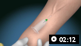
 Diagnostic lumbar puncture in adults: animated demonstration