Summary
Definition
History and exam
Key diagnostic factors
- pain
- soft-tissue swelling
- ecchymosis
- expanding hematoma
- impaired limb function
- inability to bear weight
- point tenderness
- deformity
- guarding
- wound overlying or near site of injury
- signs of vascular injury
- signs of acute compartment syndrome
- hypotension/hypovolemic shock
Other diagnostic factors
- altered nerve sensation
- impaired motor function
- bony crepitus
- callus
- reproduction of symptoms in stress fractures of the neck or shaft of the femur
Risk factors
- direct trauma
- indirect trauma
- osteoporosis (insufficiency fractures)
- chronic renal failure
- bone tumor (pathologic fractures)
- age >70 years
- age <30 years
- diabetes mellitus
- male sex (acute fractures)
- female sex (fatigue and insufficiency fractures)
- prolonged corticosteroid use (insufficiency fractures)
- low body mass index (insufficiency fractures)
- history of recent fall (insufficiency fractures)
- prior fracture (insufficiency fractures)
- seizures (proximal humerus fracture)
- long-term bisphosphonate use
- smoking
- alcohol
Diagnostic tests
1st tests to order
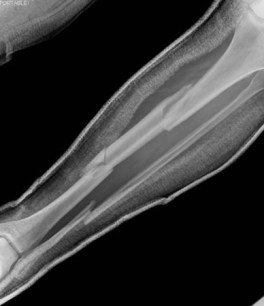
- x-ray limb
- CBC, blood typing, and cross-matching (major trauma)
- lactate
- base excess
Tests to consider
- MRI of area of interest without intravenous contrast
- noncontrast CT of fracture
- whole body bone scan with single photon emission CT (SPECT) or SPECT/CT area of interest
- compartment pressure testing
- Doppler pressure (ankle/brachial systolic pressure index)
- ultrasound duplex scanning
- CT angiogram
- angiography
- dual-energy x-ray absorptiometry bone density scan
- whole-body CT
Treatment algorithm
involved in high-energy trauma
distal humeral shaft: nonstress
midshaft humeral: nonstress
proximal humeral shaft: nonstress
radial or ulnar shaft: nonstress
upper limb stress fractures
femoral shaft: nonstress
tibia or fibula shaft: nonstress
femoral stress fractures
fibular or tibial stress fractures
Contributors
Expert advisers
Nathan Olszewski, MD
Assistant Professor of Orthopedic Surgery
Orthopedic Surgeon
Boston University Chobanian & Avedisian School of Medicine
Boston Medical Center
Department of Orthopaedic Surgery
Boston, MA
Disclosures
NO has been reimbursed for travel from Stryker Corporation for participation in educational case conferences and equipment training sessions.
Paul Tornetta, III, MD, PhD
Chief and Chair, Professor and Residency Program Director, Director of Orthopedic Trauma
Boston University Chobanian & Avedisian School of Medicine
Boston Medical Center
Department of Orthopaedic Surgery
Boston, MA
Disclosures
PT holds intellectual property with Smith & Nephew and has received publishing royalties from Wolters Kluwer.
Acknowledgements
Dr Nathan Olszewski and Dr Paul Tornetta III would like to gratefully acknowledge Dr Philip H. Cohen, the previous contributor to this topic.
Disclosures
PHC has given lectures for MCE Conferences, a medical education company, and received a stipend/free hotel room during the conference. MCE Conferences accepts no funding from pharmaceutical companies or other outside agencies, and PHC declares that the lectures have no impact on the topic.
Peer reviewers
Robert D. Golden, MD
Chief, Orthopaedic Surgery
MedStar Washington Hospital Center
Regional Chief
Orthopaedic Trauma Surgery
MedStar Orthopaedic Institute
Washington, DC
Disclosures
RDG declares that he has no competing interests.
Peer reviewer acknowledgements
BMJ Best Practice topics are updated on a rolling basis in line with developments in evidence and guidance. The peer reviewers listed here have reviewed the content at least once during the history of the topic.
Disclosures
Peer reviewer affiliations and disclosures pertain to the time of the review.
References
Key articles
American College of Radiology. ACR appropriateness criteria: stress (fatigue-insufficiency) fracture including sacrum excluding other vertebrae. 2024 [internet publication].Full text
National Institute for Health and Care Excellence. Fractures (complex): assessment and management. Nov 2022 [internet publication].Full text
American Academy of Orthopaedic Surgeons. Prevention of surgical site infections after major extremity trauma: evidence-based clinical practice guideline. Mar 2022 [internet publication].Full text
Reference articles
A full list of sources referenced in this topic is available to users with access to all of BMJ Best Practice.
Differentials
- Contusion
- Anterior glenohumeral dislocation
- Rotator cuff injury
More DifferentialsGuidelines
- ACR appropriateness criteria: stress (fatigue-insufficiency) fracture including sacrum excluding other vertebrae
- Fractures (complex): assessment and management
More Guidelines Log in or subscribe to access all of BMJ Best Practice
Use of this content is subject to our disclaimer