
Summary
Ptosis, or blepharoptosis, refers to the drooping or downward displacement of the upper eyelid. The levator muscle, its aponeurosis, and the superior tarsal muscle are responsible for upper eyelid resting position and elevation. When these structures are compromised, the resultant depressed eyelid position can reduce the amount of light entering the eye, thereby degrading visual acuity. In pseudoptosis, aberrant structural relationships of the intact globe, bony, and soft-tissue attachments may cause secondary eyelid abnormalities.
Congenital myogenic, acquired aponeurotic, and involutional forms of ptosis represent the most common causes of ptosis among children and adults.[1][2] Adults may be affected by associated involutional changes to the facial soft tissues that exacerbate or mask signs of ptosis. The vast majority of patients with ptosis do not present to the ophthalmologist or oculoplastic surgeon for evaluation and treatment. Of those who do, symptoms include headache, brow ache, and decreased visual acuity and visual field. Visual acuity improves with manual elevation of the eyelid and facial soft tissues. Superior visual field loss is most common; however, central vision can also be adversely affected. Any acute onset of ptosis, especially with other ocular or orbital symptoms, justifies further investigation with ophthalmologic consultation.[3][Figure caption and citation for the preceding image starts]: Sagittal view of eyelid anatomyFrom the collection of Dr Allen Putterman [Citation ends]. [Figure caption and citation for the preceding image starts]: Bilateral, asymmetric, congenital myogenic ptosisMid Essex Hospital Services NHS Trust/Science Photo Library; used with permission [Citation ends].
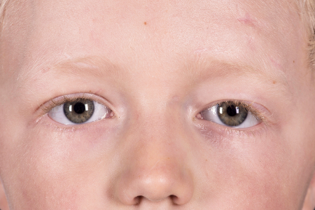 [Figure caption and citation for the preceding image starts]: Ptosis in a 6-year-old boy. Ptosis is normally due to weakness of the levator muscle of the upper eyelid, here of the left eye (at right). This patient has had this condition since birth, and has had three operations aimed at correcting the conditionMid Essex Hospital Services NHS Trust/Science Photo Library; used with permission [Citation ends].
Differentials
Common
- Involutional changes
- Prolapsed orbital fat
- Dermatochalasis
- Congenital myogenic ptosis
- Thyroid eye disease
- Previous eye-related surgery or implant
- Chalazion
- Stye (hordeolum)
- Uveitis
- Orbital cellulitis
- Orbital inflammatory syndrome
- Eyelid tumors
- Orbital tumors
- Chronic progressive external ophthalmoplegia (CPEO)
- Stroke
- Eyelid foreign body
- Eyelid laceration
Uncommon
- Blepharophimosis
- Myasthenia gravis
- Multiple sclerosis
- Blepharochalasis
- Giant cell arteritis
- Preseptal cellulitis
- Globe malposition
- Benign essential blepharospasm (BEB)
- Third nerve palsy
- Horner syndrome
- Transection of levator muscle or aponeurosis
- Orbital and facial fracture
Contributors
Authors
Kiran Sajja, MD
Milan Eye Center
Oculoplastic Surgery
Johns Creek
GA
Disclosures
KS declares that he has no competing interests.
Acknowledgements
Dr Kiran Sajja would like to gratefully acknowledge Dr Allen M. Putterman, a previous contributor for this topic.
Disclosures
AMP declares that he has no competing interests.
Peer reviewers
Ilse Mombaerts, MD, PhD
Department of Ophthalmology
University Hospitals
Leuven Kapucijnenvoer
Leuven
Belgium
Disclosures
IM declares that she has no competing interests.
Alon Kahana, MD, PhD
Assistant Professor
Kellogg Eye Center
Assistant Professor
Ophthalmology and Visual Sciences
Comprehensive Cancer Center
University of Michigan
Ann Arbor
MI
Disclosures
AK declares that he has no competing interests.
Peer reviewer acknowledgements
BMJ Best Practice topics are updated on a rolling basis in line with developments in evidence and guidance. The peer reviewers listed here have reviewed the content at least once during the history of the topic.
Disclosures
Peer reviewer affiliations and disclosures pertain to the time of the review.
References
Key articles
Finsterer J. Ptosis: causes, presentation, and management. Aesthetic Plast Surg. 2003;27:193-204. Abstract
Yanovitch T, Buckley E. Diagnosis and management of third nerve palsy. Curr Opin Ophthalmol. 2007 Sep;18(5):373-8. Abstract
American College of Radiology. ACR Appropriateness Criteria. Orbits, vision and visual loss. 2017 [internet publication].Full text
Reference articles
A full list of sources referenced in this topic is available to users with access to all of BMJ Best Practice.
Use of this content is subject to our disclaimer