Summary
Definition
The medial and lateral menisci are shock absorbers and force distributors located between the femur and the tibia. Consequently, menisci can tear due to traumatic injury or degenerative wear (e.g., in knee joint arthritis), and can compromise force distribution across the knee joint. A meniscal tear occurs in 2 primary planes, vertical and horizontal. Vertical tears are generally the result of an acute trauma, whereas horizontal tears are typically degenerative in nature.[1] Tears can cause knee pain, swelling, limited range of motion, and catching, locking, and buckling of the knee joint. Tears may lead to degenerative, arthritic changes if not already present.
[Figure caption and citation for the preceding image starts]: Anatomical structures around the menisciCreated by BMJ Publishing Group [Citation ends].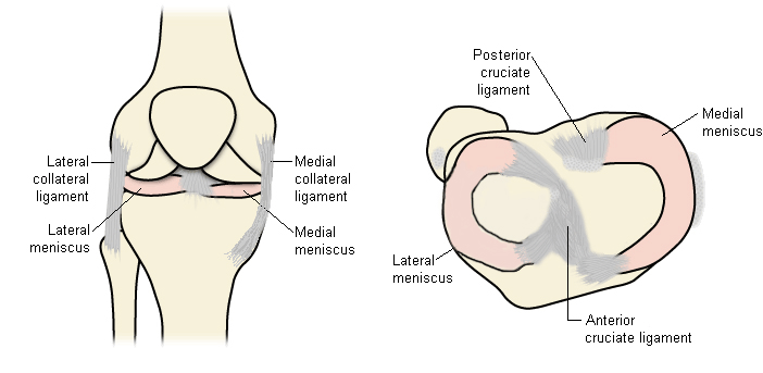
History and exam
Key diagnostic factors
- history of knee trauma
- history of knee arthritis, instability, or malalignment
- knee swelling
- sensation of knee instability or buckling/catching
- knee pain
- tenderness at joint line and joint line crepitation
Other diagnostic factors
- popliteal (Baker) cyst in chronic cases
- limited range of motion
Risk factors
- acute trauma (pivoting or twisting injury)
- knee joint arthritis
- knee instability
- history of anterior cruciate ligament injury
- malalignment of the knee joint
- rough or uneven playing surface
- poor ground/weather conditions
- older age
- construction work and manual labor jobs
- discoid meniscus
- high BMI
Diagnostic tests
1st tests to order
- clinical tests
- MRI scan
- x-ray
Tests to consider
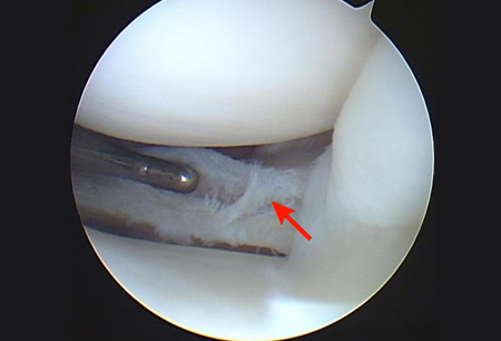
- arthroscopy
- CT arthrography
- ultrasound
Treatment algorithm
all patients
Contributors
Authors
Hideki Takeda, MD
Orthopedic Surgeon
Department of Sports Orthopedics
NTT Medical Center Tokyo
Tokyo
Japan
Disclosures
HT declares that he has no competing interests.
Acknowledgements
Dr Hideki Takeda would like to gratefully acknowledge Dr Lars Engebretsen and Dr Kevin R. Stone, previous contributors to this topic.
Disclosures
LE declares that he has no competing interests. KRS is an author of several references cited in this topic.
Peer reviewers
Daniel Solomon, MD
Co-Director of Orthopedic Sports and Shoulder Service
Department of Orthopedic Surgery
Naval Medical Center San Diego
San Diego
CA
Disclosures
DS declares that he has no competing interests.
Jung-Ro Yoon, MD
Orthopedic Surgeon
Department of Orthopedic Surgery
Seoul Veterans Hospital
Seoul
South Korea
Disclosures
JRY declares that she has no competing interests.
Nikunj N. Trivedi, MD
Fellow
Sports Medicine and Shoulder Surgery
Stanford University
Stanford
CA
Disclosures
NNT declares that he has no competing interests.
Seth L. Sherman, MD
Associate Professor of Orthopedic Surgery
Fellowship Director
Sports Medicine and Shoulder Surgery
Stanford University
Stanford
CA
Disclosures
SLS declares that he has no competing interests.
Peer reviewer acknowledgements
BMJ Best Practice topics are updated on a rolling basis in line with developments in evidence and guidance. The peer reviewers listed here have reviewed the content at least once during the history of the topic.
Disclosures
Peer reviewer affiliations and disclosures pertain to the time of the review.
References
Key articles
Kopf S, Beaufils P, Hirschmann MT, et al. Management of traumatic meniscus tears: the 2019 ESSKA meniscus consensus. Knee Surg Sports Traumatol Arthrosc. 2020 Apr;28(4):1177-94.Full text Abstract
American Physical Therapy Association. Knee pain and mobility impairments: meniscal and articular cartilage lesions, revision 2018. 2018 [internet publication].
American College of Radiology. ACR Appropriateness Criteria® acute trauma to the knee. 2019 [internet publication].Full text
Reference articles
A full list of sources referenced in this topic is available to users with access to all of BMJ Best Practice.
Differentials
- Anterior cruciate ligament tear
- Medial collateral ligament sprain
- Posterior cruciate ligament sprain
More DifferentialsGuidelines
- Acute isolated meniscal pathology
- Management of traumatic meniscus tears: the 2019 ESSKA meniscus consensus
More Guidelines Log in or subscribe to access all of BMJ Best Practice
Use of this content is subject to our disclaimer