Summary
Definition
History and exam
Key diagnostic factors
- history of traumatic or nontraumatic cutaneous lesion
- anesthesia or severe pain over site of cellulitis
- fever
- palpitations, tachycardia, tachypnea, hypotension, and lightheadedness
- nausea and vomiting
- delirium
- crepitus
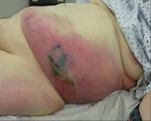
- vesicles or bullae
- gray discoloration of skin
- edema or induration
- location of lesion
Risk factors
- inpatient contact with index case
- Varicella zoster infection
- cutaneous injury, surgery, trauma
- nontraumatic skin lesions
- intravenous drug use
- chronic illness
- immunosuppression
- use of nonsteroidal anti-inflammatory drugs (NSAIDs)
Diagnostic tests
1st tests to order
- surgical exploration
- blood and tissue cultures
- gram stain
- complete blood count and differential
- serum electrolytes
- serum BUN and creatinine
- serum CRP
- serum creatine kinase (CK)
- serum lactate
- clotting screen
- arterial blood gas
Tests to consider
- radiography, CT/MRI, ultrasound
- fresh frozen section
Treatment algorithm
suspected necrotizing fasciitis, organism unknown
type I necrotizing fasciitis (polymicrobial)
type II necrotizing fasciitis due to group A streptococcus
type II necrotizing fasciitis due to Staphylococcus aureus
type II necrotizing fasciitis due to Vibrio vulnificus
type II necrotizing fasciitis due to Aeromonas hydrophila
type II necrotizing fasciitis due to mucorales
persistent cosmetic and functional defects after debridement
Contributors
Expert advisers
Ramia Zakhour, MD
Assistant Professor
Department of Pediatrics
University of Texas
McGovern Medical School
Houston, TX
Disclosures
RZ declares that they have no competing interests.
Acknowledgements
Dr Ramia Zakour would like to gratefully acknowledge Dr Kevin Steiner and Dr William Petri, previous contributors to this topic.
Disclosures
KS and WP declared they have no competing interests.
Peer reviewers
Felix Lui, MD, FACS
Associate Professor of Surgery
Yale School of Medicine
New Haven, VT
Disclosures
FL declares that he has no competing interests.
Shiranee Sriskandan, MA, MBBChir, FRCP, PhD
Professor of Infectious Diseases and Hon. Consultant
Section of Infectious Diseases
Imperial College London
London
UK
Disclosures
SS declares that she has no competing interests.
Peer reviewer acknowledgements
BMJ Best Practice topics are updated on a rolling basis in line with developments in evidence and guidance. The peer reviewers listed here have reviewed the content at least once during the history of the topic.
Disclosures
Peer reviewer affiliations and disclosures pertain to the time of the review.
References
Key articles
Sartelli M, Guirao X, Hardcastle TC, et al. 2018 WSES/SIS-E consensus conference: recommendations for the management of skin and soft-tissue infections. World J Emerg Surg. 2018 Dec 14;13:58.Full text Abstract
Stevens DL, Bisno AL, Chambers HF, et al. Practice guidelines for the diagnosis and management of skin and soft tissue infections: 2014 update by the Infectious Diseases Society of America. Clin Infect Dis. 2014 Jul 15;59(2):e10-52.Full text Abstract
Reference articles
A full list of sources referenced in this topic is available to users with access to all of BMJ Best Practice.
Differentials
- Cellulitis
- Impetigo
- Erysipelas
More DifferentialsGuidelines
- WSES/GAIS/WSIS/SIS-E/AAST global clinical pathways for patients with skin and soft tissue infections
- 2018 WSES/SIS-E consensus conference: recommendations for the management of skin and soft-tissue infections
More Guidelines Log in or subscribe to access all of BMJ Best Practice
Use of this content is subject to our disclaimer