Summary
Definition
Seborrheic keratosis is a common, benign skin tumor most commonly found on the torso and forehead. The lesions classically appear in multiples as well-circumscribed plaques with a "stuck-on" appearance. Later the plaques can become raised and may show a verrucous surface. Lesions can display a variety of colors, but are usually yellow or light-to-dark brown. Most lesions do not exceed 1 cm in diameter. The lesions are normally painless and require no treatment.[1][2][3][Figure caption and citation for the preceding image starts]: Seborrheic keratosis on the chest of an elderly womanFrom the collection of Dr Braun and Dr Kolm, used with permission [Citation ends]. [Figure caption and citation for the preceding image starts]: Seborrheic keratosis of the chest: clinical overview imageFrom the collection of Dr Braun and Dr Kolm, used with permission [Citation ends].
 [Figure caption and citation for the preceding image starts]: Seborrheic keratosis of the chest: clinical close-up imageFrom the collection of Dr Braun and Dr Kolm, used with permission [Citation ends].
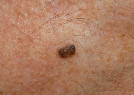 [Figure caption and citation for the preceding image starts]: Dermoscopic image of seborrheic keratosis on the chestFrom the collection of Dr Braun and Dr Kolm, used with permission [Citation ends].
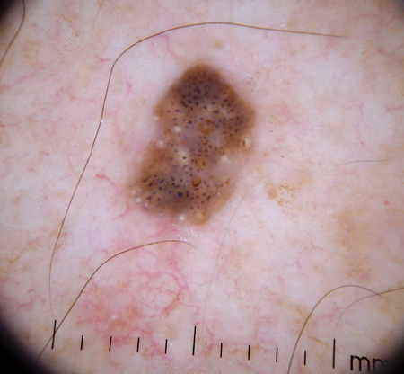 [Figure caption and citation for the preceding image starts]: Clinical close-up image of seborrheic keratosis on the back of a 40-year-old manFrom the collection of Dr Braun and Dr Kolm, used with permission [Citation ends].
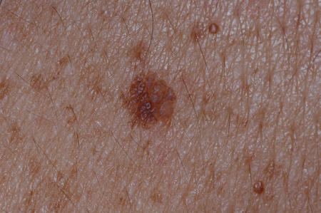 [Figure caption and citation for the preceding image starts]: Clinical overview image of seborrheic keratosis on the back of a 40-year-old manFrom the collection of Dr Braun and Dr Kolm, used with permission [Citation ends].
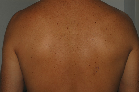 [Figure caption and citation for the preceding image starts]: Clinical image of an example of a dark-brown pigmented seborrheic keratosisFrom the collection of Dr Braun and Dr Kolm, used with permission [Citation ends].
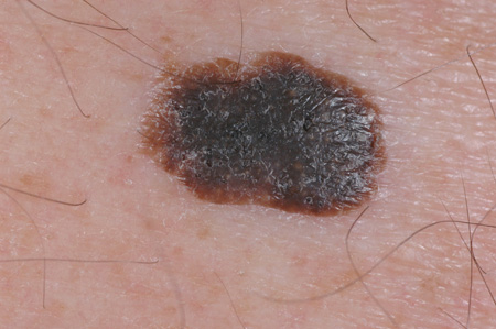 [Figure caption and citation for the preceding image starts]: Example of a dark-brown pigmented seborrheic keratosis. Dermoscopic image: see yellowish horn pearls and dark brown holes corresponding to so-called "pseudo-follicular openings"From the collection of Dr Braun and Dr Kolm, used with permission [Citation ends].
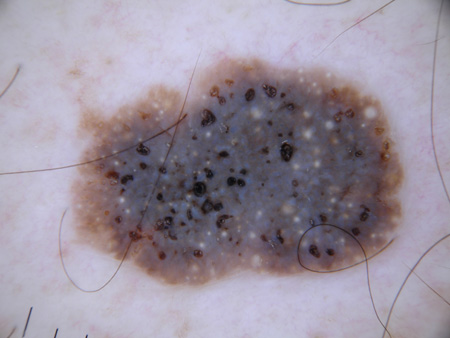
History and exam
Key diagnostic factors
- lesions appear "stuck-on"
- localization on torso or face
- yellow or light- to dark-brown-colored lesions
- slightly raised, flat surface lesions
- wart-like texture
- multiple lesions
- painless
- itching (prurigo)
Other diagnostic factors
- round yellow-white horn pearls in the surface of lesions
Risk factors
- age over 50 years
- Fitzpatrick skin type I or II
- Fitzpatrick skin type IV, V, or VI (dermatosis papulosa nigra)
- female sex (dermatosis papulosa nigra)
- family history
- sun/UV exposure
- pregnancy
Diagnostic investigations
Investigations to consider
- dermoscopy
- biopsy and histopathologic examination
- reflectance confocal microscopy (RCM)
Treatment algorithm
irritated or itching lesions
raised seborrheic keratosis
flat seborrheic keratosis
Contributors
Authors
Ralph Braun, MD
Professor
Clinic Utoquai
Zurich
Switzerland
Disclosures
RB declares that he has no competing interests.
Isabel Kolm-Djamei, MD
Consultant Dermatologist
Department of Pathology
Cantonal Hospital Lucerne
Lucerne
Switzerland
Disclosures
IKD declares that she has no competing interests.
Peer reviewers
Erin Warshaw, MD, MS
Associate Professor
Department of Dermatology
University of Minnesota
MN
Disclosures
EW declares that he has no competing interests.
Peer reviewer acknowledgements
BMJ Best Practice topics are updated on a rolling basis in line with developments in evidence and guidance. The peer reviewers listed here have reviewed the content at least once during the history of the topic.
Disclosures
Peer reviewer affiliations and disclosures pertain to the time of the review.
References
Key articles
Seaton E, Madan V. Benign keratinocytic acanthomas and proliferations. In: Barker J, Griffiths C, Bleiker T, eds. Rook's textbook of dermatology. 10th ed. Hoboken, NJ: John Wiley & Sons, Ltd; 2024.
Patterson JW. Chapter 32: Tumors of the epidermis. In: Patterson JW. Weedon's skin pathology. 6th ed. Philadelphia, PA: Elsevier; 2025: 877-936.
Barthelmann S, Butsch F, Lang BM, et al. Seborrheic keratosis. J Dtsch Dermatol Ges. 2023 Mar;21(3):265-77.Full text Abstract
Reference articles
A full list of sources referenced in this topic is available to users with access to all of BMJ Best Practice.
Differentials
- Malignant melanoma
- Viral warts
- Nevus
More Differentials Log in or subscribe to access all of BMJ Best Practice
Use of this content is subject to our disclaimer